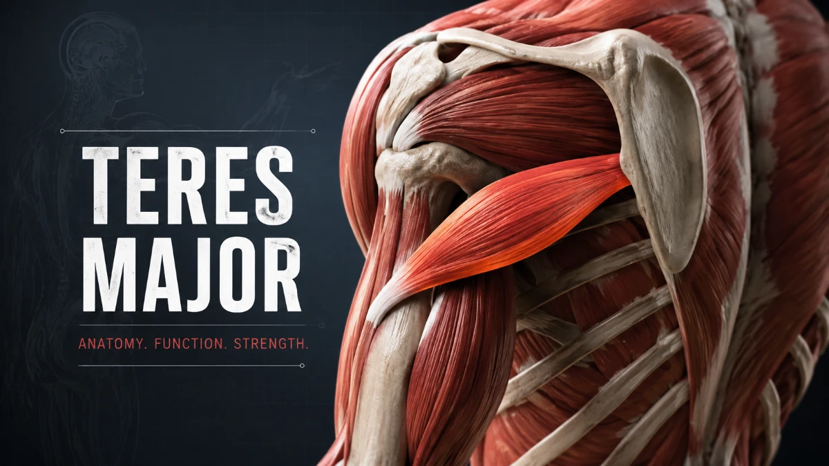
The teres major is a thick posterior shoulder muscle that runs from the lower part of the shoulder blade to the upper arm bone. It helps extend the arm backward, pull it toward the body, and rotate it inward. It works closely with the latissimus dorsi during pulling, climbing, swimming, rowing, and throwing movements, but it is not part of the rotator cuff.
Teres Major Anatomy at a Glance
| Feature | Details |
| Muscle type | Intrinsic shoulder-region muscle |
| Location | Posterior and inferior shoulder, near the back of the axilla |
| Origin | Posterior surface of the inferior angle of the scapula and adjacent lower lateral border |
| Insertion | Medial lip of the intertubercular groove of the humerus |
| Main actions | Shoulder extension, adduction, and medial rotation |
| Typical nerve supply | Lower subscapular nerve |
| Brachial plexus relationship | Usually a branch of the posterior cord |
| Functional partner | Latissimus dorsi |
| Rotator cuff member | No |
| Key anatomical relationship | Forms the inferior boundary of the quadrangular space |
What Is the Teres Major Muscle?
The teres major is a broad scapulohumeral muscle located on the lower back portion of the shoulder. It begins at the inferior angle of the scapula—the lowest point of the shoulder blade—and passes upward and laterally to the humerus.
Its line of pull explains its actions. When it contracts, it draws the upper arm backward, toward the trunk, and into medial rotation. It is active when bringing an elevated arm down against resistance, including during pull-ups, swimming pull-throughs, rowing, and climbing.
Read more: Teres Major
Teres Major Location and Surface Anatomy
This muscle lies below the teres minor and near the lower lateral border of the scapula. As it travels toward the humerus, it blends visually and functionally with the latissimus dorsi near the back of the armpit.
Together, the teres major and latissimus dorsi create much of the posterior axillary fold, the muscular border seen at the rear of the armpit.
The muscle is positioned:
- Inferior to the teres minor
- Posterior to the axilla
- Deep to parts of the deltoid
- Adjacent to the long head of the triceps brachii
- Close to the latissimus dorsi near the humeral attachment
- Inferior to the quadrangular space
This crowded anatomical region contains muscles, tendons, nerves, vessels, and joint structures. As a result, pain near the posterior shoulder or axilla cannot be attributed to this muscle on location alone.
Teres Major Origin and Insertion
The teres major has a broad scapular attachment and a comparatively short distal tendon.
Origin
The muscle originates from:
- The posterior surface of the inferior angle of the scapula
- The adjacent lower portion of the lateral border of the scapula
This location places it below the teres minor and near the posterior shoulder structures that guide movement around the glenohumeral joint.
Insertion
The tendon inserts on the medial lip of the intertubercular groove of the humerus, also called the crest of the lesser tubercle.
The intertubercular groove, commonly known as the bicipital groove, houses the tendon of the long head of the biceps brachii. Three major muscles attach around it:
| Muscle | Attachment around the intertubercular groove |
| Pectoralis major | Lateral lip |
| Latissimus dorsi | Floor |
| Teres major | Medial lip |
This arrangement helps explain the muscle’s ability to adduct and medially rotate the humerus.
Teres Major Function
The teres major acts mainly at the glenohumeral joint. Its three primary actions are:
- Shoulder extension: moves the upper arm backward
- Shoulder adduction: draws the arm toward the side of the body
- Medial rotation: turns the humerus inward
It commonly works with the latissimus dorsi during forceful pulling movements. The two muscles are especially important when the arm moves from an overhead position toward the trunk.
Activities That Use the Teres Major
This muscle contributes during:
- Pull-ups and lat pulldowns
- Rowing and paddling
- Rope climbing
- Swimming pull-throughs
- Gymnastics
- Throwing and racket sports
- Pulling a heavy object toward the torso
- Bringing the arm down from an overhead position
The muscle rarely works in isolation. These movements also require coordinated activity from the scapular stabilizers, rotator cuff, deltoid, triceps, and other upper-body muscles.
Teres Major Nerve Supply
The teres major is usually innervated by the lower subscapular nerve, a branch of the posterior cord of the brachial plexus. The nerve commonly carries fibers from C5 to C7, although root-level descriptions may vary among anatomy texts.
The lower subscapular nerve also supplies the inferior portion of the subscapularis. Therefore, injury involving the posterior cord or lower subscapular nerve can affect more than one shoulder muscle.
Anatomical variation can occur. In a minority of people, the thoracodorsal nerve may contribute to the nerve supply of the muscle. This variation has practical relevance in reconstructive procedures and tendon-transfer surgery.
Blood Supply
Arterial supply usually comes from branches of the subscapular artery system, including thoracodorsal and circumflex scapular branches. Individual vascular patterns can vary, which is relevant in surgical planning.
Teres Major vs Teres Minor vs Latissimus Dorsi
The teres major is commonly confused with the teres minor because of their similar names and proximity. It also has a close functional relationship with the latissimus dorsi.
| Feature | Teres major | Teres minor | Latissimus dorsi |
| Primary location | Lower posterior shoulder | Posterior shoulder, above teres major | Broad back and posterior axillary fold |
| Origin | Inferior angle and lower lateral border of scapula | Lateral border of scapula | Thoracolumbar fascia, lower thoracic region, iliac crest, and lower ribs |
| Insertion | Medial lip of intertubercular groove | Inferior facet of greater tubercle | Floor of intertubercular groove |
| Main action | Extension, adduction, medial rotation | External rotation and shoulder stabilization | Extension, adduction, medial rotation |
| Primary nerve | Lower subscapular nerve | Axillary nerve | Thoracodorsal nerve |
| Rotator cuff muscle? | No | Yes | No |
| Functional relationship | Works closely with latissimus dorsi | Works with infraspinatus | Works closely with teres major |
The teres minor is a rotator cuff muscle and helps externally rotate the shoulder. By contrast, the teres major performs medial rotation and is functionally more similar to the latissimus dorsi.
Is the Teres Major Part of the Rotator Cuff?
No. The teres major is not a rotator cuff muscle.
The rotator cuff includes:
- Supraspinatus
- Infraspinatus
- Teres minor
- Subscapularis
These muscles attach around the humeral head and help stabilize it in the glenoid during arm movement. The teres major inserts at the intertubercular groove and acts mainly as an extensor, adductor, and medial rotator of the upper arm.
Relationship to the Quadrangular Space
The muscle forms the inferior boundary of the quadrangular space, an anatomical passageway in the posterior shoulder.
The quadrangular space is bounded by:
- Superiorly: teres minor
- Inferiorly: teres major
- Medially: long head of the triceps brachii
- Laterally: surgical neck of the humerus
The axillary nerve and posterior circumflex humeral artery pass through this space. This relationship matters in posterior shoulder trauma, surgery, and conditions involving the axillary nerve.
Teres Major Pain and Injury Symptoms
Injury to this muscle is less common than rotator cuff tendinopathy, but it can occur during powerful overhead, traction, or pulling movements. It may be injured alone or together with the latissimus dorsi because the muscles work together and have closely related anatomy.
Common Injury Mechanisms
Potential causes include:
- High-velocity throwing, particularly pitching
- Water-skiing, climbing, or sudden traction on the arm
- Heavy pulling exercises
- Swimming and rowing
- Racket sports
- Sudden eccentric loading while the arm is overhead
- Falls or direct trauma to the posterior shoulder
Symptoms That May Occur
A strain or tear may cause:
- Pain at the lower back of the shoulder
- Tenderness near the posterior axillary fold
- Pain during pulling, throwing, or lowering the arm from overhead
- Weakness with resisted shoulder extension, adduction, or medial rotation
- Swelling or bruising after an acute injury
- Reduced force during sport-specific movements
These symptoms are not specific. Similar complaints can arise from the latissimus dorsi, rotator cuff, long-head biceps tendon, triceps, cervical spine, brachial plexus, or the glenohumeral joint.
Muscle Soreness, Trigger Points, and Injury
The phrase “trigger point” is often used to describe a tender point in a muscle that is painful when pressed. However, tenderness near the posterior shoulder or axilla cannot reliably identify the exact pain source.
Pain in this region may be felt around:
- The lower posterior shoulder
- The posterior armpit
- The upper arm
- The shoulder during pulling or overhead movement
Delayed-onset muscle soreness after unfamiliar exercise usually develops gradually and improves over several days. Sudden sharp pain, a popping sensation, bruising, clear weakness, or persistent symptoms after a throwing or pulling injury should not be assumed to be muscle tightness.
How Clinicians Evaluate a Suspected Injury
No single home test can confirm an injury to this muscle. Clinicians use the mechanism of injury, physical examination, strength testing, and imaging when needed.
History and Inspection
A clinician may ask about:
- Whether pain began suddenly or developed gradually
- The exact movement that triggered symptoms
- Recent throwing, swimming, climbing, or resistance-training load
- A pop, bruising, swelling, or immediate weakness
- Previous shoulder or neck injuries
- Tingling, numbness, or altered sensation
The posterior axillary fold may be examined for swelling, bruising, asymmetry, or changes in contour after a significant injury.
Movement and Strength Testing
Because this muscle helps extend, adduct, and medially rotate the shoulder, a clinician may assess pain and strength during resisted versions of those movements.
These tests are not completely specific. They also recruit the latissimus dorsi, pectoralis major, subscapularis, posterior deltoid, and other muscles. Clinicians interpret test findings alongside tenderness, shoulder range of motion, scapular control, neurological signs, and the mechanism of injury.
Palpation Considerations
The teres major lies within a dense anatomical area near the latissimus dorsi, triceps, scapular muscles, and important nerves and vessels. Tenderness near the posterior axillary fold can support clinical suspicion, but palpation alone cannot distinguish an injury from nearby soft-tissue conditions.
MRI and Ultrasound Findings
MRI and ultrasound can help assess suspected muscle or tendon injury, particularly when symptoms began after trauma, bruising is present, strength loss is substantial, or the patient is an athlete returning to high-demand sport.
Possible findings include:
- Muscle edema in an acute strain
- Injury at the myotendinous junction
- Partial muscle or tendon tearing
- Full-thickness tear
- Tendon discontinuity or retraction
- Chronic muscle atrophy or fatty change after longstanding injury or denervation
A routine shoulder MRI may not include the entire muscle, myotendinous junction, posterior axilla, and humeral insertion. When injury to this area is suspected, the clinician should communicate that concern so the scan can include the relevant anatomy.
Teres Major Injury vs Rotator Cuff Injury
Pain pattern and mechanism can guide clinical suspicion, but they cannot establish a diagnosis without an appropriate examination.
| Feature | Teres major or latissimus dorsi injury | Rotator cuff condition |
| Typical pain area | Posterior armpit or lower back of shoulder | Lateral or anterior shoulder, though patterns vary |
| Common provoking activity | Forceful throwing, pulling, climbing, water-skiing | Repetitive overhead use, lifting, falls, tendon degeneration |
| Main painful actions | Extension, adduction, medial rotation, powerful pull-through | Elevation, external rotation, resisted abduction, overhead reach |
| Possible acute findings | Bruising, posterior axillary tenderness, reduced pulling force | Painful arc, night pain, weakness with abduction or external rotation |
| Imaging focus | Muscle belly, myotendinous junction, humeral insertion | Supraspinatus, infraspinatus, subscapularis, biceps tendon, subacromial structures |
The patterns in this table are broad clinical clues, not diagnostic rules. Symptoms can overlap, and more than one shoulder condition may be present.
Recovery and Return to Activity
Recovery depends on whether symptoms arise from overload, a mild strain, a partial tear, a tendon injury, or another shoulder condition. A return-to-sport decision should be based on function rather than a fixed calendar timeline.
Mild Overload or Grade I Strain
A mild strain is often managed by temporarily reducing painful activity while maintaining comfortable motion. Strengthening can gradually resume as pain settles and the shoulder tolerates progressive loading.
Moderate Muscle or Myotendinous Injury
A more substantial injury may require a structured rehabilitation plan. Early management typically protects painful tissue while preserving range of motion. Progressive rehabilitation then restores shoulder strength, scapular control, pulling tolerance, and sport-specific capacity.
Severe Tear or Tendon Injury
A complete tear, visible change in the posterior axillary fold, marked weakness, or injury in a high-demand throwing athlete should be assessed by a sports medicine or orthopedic specialist. Treatment may be nonoperative or surgical depending on the injury pattern, tendon retraction, functional demands, and individual goals.
Return-to-Sport Checklist
Before returning to throwing, climbing, swimming, or heavy pulling, an individual should generally have:
- Functional, comfortable shoulder range of motion
- No significant pain with resisted extension, adduction, or medial rotation
- Strength adequate for the demands of the activity
- Tolerance of progressively harder training sessions
- No substantial next-day symptom flare after sport-specific loading
- Clearance from a treating clinician after a significant tear or persistent symptoms
Surgical Relevance
The teres major has a role in selected reconstructive shoulder procedures and tendon transfers. In some cases of severe, irreparable rotator cuff deficiency, surgeons may consider tendon transfer to restore a lost movement or improve shoulder function.
Its relationship with the latissimus dorsi, blood supply, nerve entry points, and nearby neurovascular structures matter during surgical planning. These specialized procedures are not routine treatments for ordinary shoulder pain or a simple muscle strain.
When to Seek Medical Care
Seek prompt medical assessment after a forceful throwing or pulling injury, significant trauma, or a fall when symptoms include:
- A pop at the time of injury
- Extensive bruising or swelling
- A visible change near the posterior shoulder or armpit
- Marked weakness or inability to pull, lift, or throw
- Numbness, tingling, or new sensory changes
- A shoulder that appears dislocated
- A cold, pale, or unusually swollen arm
- Pain that persists or worsens despite activity modification
Key Takeaways
The teres major is a posterior shoulder muscle that extends from the inferior scapula to the medial lip of the humeral intertubercular groove. It extends, adducts, and medially rotates the arm.
It is usually supplied by the lower subscapular nerve, functions closely with the latissimus dorsi, and forms the inferior boundary of the quadrangular space. Although it lies near the teres minor, it is not part of the rotator cuff.
Pain or injury in this area can resemble other shoulder disorders. Sudden posterior shoulder or armpit pain with bruising, weakness, or reduced athletic performance merits professional assessment.
Frequently Asked Questions
Where is the teres major muscle located?
The teres major is located on the lower back portion of the shoulder. It starts from the inferior angle and lower lateral border of the scapula, then travels toward the humerus near the posterior axilla. Together with the latissimus dorsi, it forms much of the posterior axillary fold. Its position below the teres minor and near the long head of the triceps helps identify it within posterior shoulder anatomy.
What are the three main actions of the teres major?
Its three main actions are shoulder extension, adduction, and medial rotation. Extension moves the upper arm backward, adduction brings it toward the body, and medial rotation turns the humerus inward. These actions support pulling and downward arm movements, including rowing, climbing, swimming, pull-ups, and certain phases of throwing.
What nerve supplies the teres major?
The lower subscapular nerve usually supplies this muscle. It arises from the posterior cord of the brachial plexus and commonly contains nerve fibers from C5 to C7. In a minority of individuals, the thoracodorsal nerve may contribute to its innervation. This variation matters in procedures around the posterior axilla and in reconstructive tendon-transfer surgery.
Is the teres major part of the rotator cuff?
No. The teres major is separate from the rotator cuff. The rotator cuff consists of the supraspinatus, infraspinatus, teres minor, and subscapularis. Teres major attaches to the medial lip of the intertubercular groove and helps extend, adduct, and internally rotate the arm. Teres minor attaches to the greater tubercle and contributes to external rotation and shoulder stabilization.
How can teres major pain be distinguished from rotator cuff pain?
Pain from the teres major or latissimus dorsi is often felt near the posterior armpit or lower back of the shoulder after forceful pulling or throwing. Rotator cuff problems more often cause lateral or anterior shoulder pain and may be aggravated by elevation or external rotation. However, pain patterns overlap, so location alone cannot identify the involved tissue. Clinical examination and, when necessary, imaging help clarify the diagnosis.
What does a teres major strain feel like?
A strain may cause pain or tenderness at the lower posterior shoulder or posterior axilla, especially during pulling, throwing, swimming, or lowering the arm from overhead. Symptoms can increase with resisted shoulder extension, adduction, or medial rotation. More serious injuries may cause sharp pain, a popping sensation, bruising, swelling, or weakness. Those signs should be evaluated rather than treated as routine post-exercise soreness.
Can an MRI show a teres major tear?
Yes. MRI can show muscle edema from a strain, injury at the myotendinous junction, partial tearing, complete tendon disruption, retraction, and chronic muscle changes. A standard shoulder MRI may not cover the full posterior axilla or the muscle’s humeral insertion. If a teres major or latissimus dorsi injury is suspected, the clinician can request imaging that includes the relevant anatomy.
How long does a teres major injury take to heal?
Healing time varies according to whether the problem is overload, a mild strain, partial tear, or tendon injury. Return to activity should depend on comfortable movement, adequate strength, and tolerance of progressive sport-specific loading rather than a fixed number of days or weeks. Sudden pain with bruising, weakness, or visible contour changes should be assessed promptly because it may indicate a more substantial injury.
Can you palpate the teres major muscle?
A clinician may palpate this region near the posterior axillary fold, particularly when shoulder position makes the muscle more prominent. However, it lies close to the latissimus dorsi, triceps, scapular muscles, nerves, and vessels. Tenderness in that location does not confirm which tissue is responsible. Palpation should be interpreted with the injury history, movement testing, strength findings, and imaging when appropriate.
Why are the teres major and latissimus dorsi often injured together?
The teres major and latissimus dorsi frequently work together because both extend, adduct, and medially rotate the arm. They also lie close to one another near the posterior axilla and humeral attachment. High-force throwing, water-skiing, climbing, and pulling can load both muscles at the same time. An injury may therefore involve one muscle, a myotendinous junction, or the nearby combined tendon region.