Last updated: August 17, 2026
Sources reviewed: NCBI Bookshelf/StatPearls, peer-reviewed anatomy and orthopaedic literature, and reputable anatomy references.
Medical review: This educational article has not been individually medically reviewed. It is not a substitute for diagnosis or personalised medical advice.
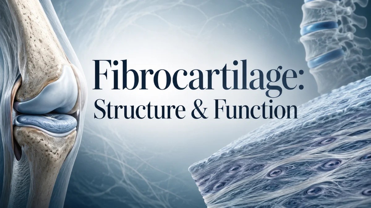
Fibrocartilage is a specialized type of cartilage adapted to areas exposed to substantial mechanical stress. It combines characteristics of dense fibrous connective tissue with those of cartilage, helping it tolerate tension, compression, and shear.
It contains abundant type I collagen along with type II collagen, relatively little ground substance, and fewer proteoglycans than hyaline cartilage. This collagen-rich structure helps fibrocartilage withstand repeated loading and tensile forces while retaining some resistance to compression
Common examples include the knee menisci, intervertebral discs, pubic symphysis, glenoid and acetabular labra, triangular fibrocartilage complex (TFCC), and specialized entheses. Unlike hyaline and elastic cartilage, fibrocartilage has no perichondrium. Its exact composition and mechanical behavior vary by anatomical location and the forces placed on it.
Fibrocartilage at a Glance
| Feature | Fibrocartilage |
| Main collagen | Type I, with type II also present |
| Main cells | Chondrocytes and fibroblast-like cells |
| Matrix | Dense, collagen-rich matrix with relatively low proteoglycan content |
| Perichondrium | Absent |
| Mechanical role | Resists tension, compression, and shear |
| Common locations | Menisci, intervertebral discs, labra, pubic symphysis, TFCC, specialized entheses |
| Healing | Generally slow; depends on the structure, vascularity, injury, and loading environment |
What Is Fibrocartilage Made Of?
The structure of fibrocartilage is closely related to its mechanical function. Its extracellular matrix contains densely arranged collagen fibers, particularly type I collagen. Type II collagen is also present, although the relative proportions and arrangement differ between tissues
Compared with hyaline cartilage, fibrocartilage contains less proteoglycan-rich ground substance. The resulting matrix is dense and suited to areas that must repeatedly tolerate mechanical forces
Its principal structural components are:
- Type I collagen: Provides substantial tensile strength.
- Type II collagen: Supports cartilage-matrix organization and resistance to compression.
- Chondrocytes: Cartilage cells found within lacunae.
- Fibroblast-like cells: Contribute to the tissue’s fibrous character.
- Extracellular matrix: The collagen- and proteoglycan-containing framework that distributes mechanical loading.
Fibrocartilage is not compositionally identical everywhere. At fibrocartilaginous entheses, for example, uncalcified fibrocartilage contains aggrecan and collagens I–III, while calcified fibrocartilage is relatively rich in type II collagen
Read more: Fibrocartilage
Fibrocartilage Histology
Under a microscope, fibrocartilage typically appears as a dense collagen-rich tissue with relatively few cells. Chondrocytes lie in lacunae between thick collagen bundles and may appear in rows, helping distinguish fibrocartilage from the smoother, more uniform matrix of hyaline cartilage
| Histological feature | Fibrocartilage |
| Collagen | Type I prominent; type II also present |
| Cells | Chondrocytes and fibroblast-like cells |
| Lacunae | Chondrocytes located between collagen bundles |
| Matrix | Dense and collagen-rich |
| Ground substance | Relatively limited compared with hyaline cartilage |
| Perichondrium | Absent |
| Overall appearance | Thick collagen bundles with relatively few cells |
What Is the Function of Fibrocartilage?
The main function of fibrocartilage is to withstand and distribute mechanical forces. Its collagen-rich architecture lets it tolerate tension and compression while helping absorb or redistribute loads
Depending on its location, fibrocartilage can:
- Resist compression
- Resist tensile forces
- Help distribute mechanical loads
- Reduce concentrated stress
- Contribute to joint stability
- Provide cushioning between structures
- Help tissues tolerate repetitive motion and friction
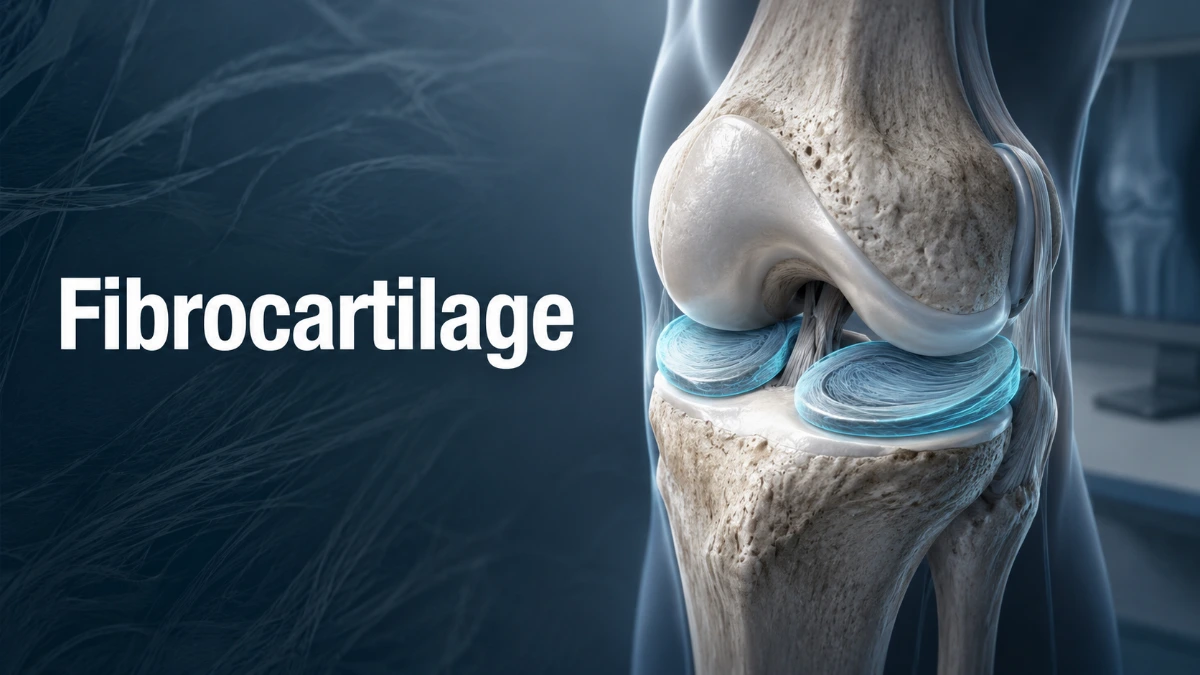
The knee menisci help distribute loads through the knee. Fibrocartilage in intervertebral discs helps the spine tolerate and distribute force. The glenoid and acetabular labra deepen their joint sockets and contribute to stability.
Fibrocartilage is therefore more than a passive cushion: it is adapted to manage force transfer between tissues and structures exposed to different types of loading.
Where Is Fibrocartilage Found?
Fibrocartilage occurs mainly where the body needs a combination of strength, cushioning, load distribution, and resistance to mechanical stress.
| Location | Main role |
| Knee menisci | Distribute loads and improve joint mechanics |
| Intervertebral discs | Help distribute spinal loads and resist deformation |
| Pubic symphysis | Helps transfer forces between the pubic bones |
| Glenoid labrum | Deepens the shoulder socket and supports stability |
| Acetabular labrum | Deepens the hip socket and contributes to stability |
| Temporomandibular joint | Articular tissue adapted to repeated loading |
| TFCC | Contributes to wrist load transmission and stability |
| Specialized entheses | Helps manage the transition between soft tissue and bone |
Knee Menisci
The menisci are fibrocartilaginous structures in the knee. They distribute loads, improve joint mechanics, and reduce concentrated stress. Their blood supply varies by region, which affects the healing potential of a tear
Intervertebral Discs
Fibrocartilaginous tissue is an important component of intervertebral discs, particularly the annulus fibrosus. The annulus helps contain the disc and distribute forces between vertebral bodies, helping the spine resist deformation under load.
Pubic Symphysis
The pubic symphysis is a fibrocartilaginous joint between the two pubic bones. Its structure helps the pelvis tolerate and transfer mechanical forces.
Glenoid and Acetabular Labra
The glenoid labrum of the shoulder and acetabular labrum of the hip are fibrocartilaginous structures that deepen their respective joint sockets. This improves the fit between joint surfaces and contributes to joint stability.
Temporomandibular Joint
The temporomandibular joint (TMJ) contains fibrocartilaginous articular tissue adapted to repeated mechanical loading from jaw movement.
Triangular Fibrocartilage Complex
The triangular fibrocartilage complex, or TFCC, is a fibrocartilage-containing structure in the wrist that contributes to load transmission and stability. It can be injured after a fall on an outstretched hand, repetitive loading, or rotational stress.
Fibrocartilaginous Entheses
Some tendon- and ligament-to-bone attachment sites contain fibrocartilage. At these entheses, fibrocartilage creates a gradual transition between soft tissue and mineralized bone, helping manage differences in stiffness and reduce stress concentration
Does Fibrocartilage Have a Perichondrium?
No. Fibrocartilage does not have a perichondrium . This distinguishes it from most hyaline and elastic cartilage, though articular hyaline cartilage is also an important exception because it lacks a perichondrium.
The absence of a perichondrium is relevant to repair, but it is not the only factor. Healing also depends on local vascularity, anatomy, injury type, and mechanical environment.
What Are the Types of Fibrocartilage?
Fibrocartilage is one of the three major cartilage categories, alongside hyaline cartilage and elastic cartilage. Some anatomy references additionally describe fibrocartilage by anatomical or functional patterns. These labels are descriptive rather than universally standardized biological subtypes.
1. Intra-Articular Fibrocartilage
Intra-articular fibrocartilage occurs inside joints and is exposed to repeated loading and movement. The knee menisci are a major example; they distribute load and reduce concentrated stress.
2. Connecting Fibrocartilage
Connecting fibrocartilage occurs in structures that connect or separate bones while helping distribute mechanical forces. Intervertebral discs are the classic example.
3. Stratified Fibrocartilage
Stratified fibrocartilage forms a thin layer in areas where tendons glide or change direction over bone. It is described in areas associated with tendons such as the tibialis posterior and peroneus longus, where it helps tissues tolerate pressure and repetitive loading.
4. Circumferential Fibrocartilage
Circumferential fibrocartilage occurs in ring-shaped structures around joint margins. The glenoid and acetabular labra are examples; they deepen joint sockets and contribute to stability.
Fibrocartilage vs. Hyaline Cartilage vs. Elastic Cartilage
Fibrocartilage, hyaline cartilage, and elastic cartilage are the three major cartilage categories, but each is adapted to a different mechanical environment.
| Feature | Fibrocartilage | Hyaline cartilage | Elastic cartilage |
| Main collagen/fibers | Type I and type II collagen | Predominantly type II collagen | Type II collagen plus elastic fibers |
| Perichondrium | Absent | Usually present; absent at articular surfaces | Present |
| Proteoglycan content | Relatively low | Relatively high | Moderate |
| Tensile strength | High | Moderate | Lower |
| Flexibility | Low to moderate | Moderate | Highest |
| Matrix appearance | Dense collagen bundles | Smooth, glassy matrix | Elastic-fiber-rich matrix |
| Typical locations | Menisci, discs, labra, pubic symphysis | Articular surfaces, nose, trachea, growth plates | External ear, epiglottis, parts of larynx |
| Main role | Resists tension, compression, and shear | Smooth support and low-friction movement | Flexible structural support |
The key difference is specialization: fibrocartilage is adapted to substantial mechanical loading; hyaline cartilage provides smooth, resilient support and low-friction articulation; and elastic cartilage is adapted for greater flexibility.
Why Is Fibrocartilage So Strong?
Fibrocartilage is adapted to high mechanical loading because of its dense collagen network, especially its abundance of type I collagen. Type I collagen resists tensile force, while the cartilage matrix and proteoglycans help the tissue tolerate compression .
Many fibrocartilaginous structures experience tension and compression at the same time. It is therefore more precise to say that fibrocartilage is the cartilage type especially adapted to substantial tensile and mechanical loading, rather than simply calling it the strongest cartilage in every possible sense.
Is Fibrocartilage Vascular or Avascular?
Cartilage tissue is generally avascular, and nutrients reach many cartilage cells largely by diffusion from surrounding tissues. However, fibrocartilage-containing structures do not all have identical blood supplies.
The knee meniscus is a useful example. Its outer region has better blood supply than its inner region, and repair potential generally falls as vascularity decreases toward the centre
For this reason, describing every fibrocartilage-containing structure as completely avascular is too simplistic. Vascularity and healing potential depend on the structure and its anatomical location.
Why Does Fibrocartilage Heal Slowly?
Fibrocartilage often heals slowly because of limited direct blood supply, relatively slow matrix turnover, and the need to repair tissue that remains exposed to mechanical loading. Nutrient delivery largely occurs through diffusion, which can limit the speed and extent of repair
Healing also differs substantially between structures. In the meniscus, tears in the more vascular outer region generally have greater repair potential than tears in the relatively avascular inner region
Factors that influence healing include:
- Blood supply
- Anatomical location
- Injury severity
- Mechanical environment
- Extent of tissue damage
There is no single healing pattern for every fibrocartilage-containing tissue.
Common Fibrocartilage-Related Injuries
Fibrocartilage-containing structures can be affected by trauma, repetitive loading, degeneration, and altered joint mechanics.
Meniscus Injuries
Meniscal tears are common knee injuries. Because vascularity differs across the meniscus, healing potential depends significantly on where a tear occurs. Tears closer to the vascular outer region generally have greater repair potential than tears in the relatively avascular inner region
Intervertebral Disc Degeneration
Fibrocartilaginous intervertebral discs help the spine distribute and withstand force. Degenerative changes can alter disc composition, water content, proteoglycans, collagen organization, and mechanical behavior.
Labral Injuries
The glenoid and acetabular labra contribute to joint stability and socket depth. They can be injured through trauma, repetitive loading, or joint instability. At the shoulder, glenoid-labral injury is clinically relevant in lesions such as a Bankart lesion.
Bankart and Hill-Sachs lesions are both associated with shoulder instability, but they do not necessarily occur together in every person.
TFCC Injuries
The TFCC contributes to wrist load transmission and stability. It may be injured after a fall on an outstretched hand or by rotational stress. Limited blood supply in some portions can make healing more difficult in certain injuries.
Can Fibrocartilage Regenerate After Injury?
Some fibrocartilage-containing tissues can repair, but the extent of healing is limited and depends heavily on the specific structure and injury. A region with better vascularity may have greater healing potential than a poorly vascularized region
Important factors include blood supply, anatomical location, injury severity, mechanical environment, and the extent of tissue damage. Fibrocartilage healing should therefore be considered structure by structure, not as one uniform process.
Is Fibrocartilage the Strongest Type of Cartilage?
Fibrocartilage is commonly considered the cartilage type best adapted to substantial tensile and mechanical loading because of its dense type I collagen network. But “strongest” does not mean best suited for every function.
Hyaline cartilage is better suited to smooth, low-friction articulation and resilient support. Elastic cartilage is specialized for greater flexibility. Each cartilage category is adapted to a distinct mechanical environment.
Key Takeaways About Fibrocartilage
- Fibrocartilage is a collagen-rich cartilage type specialized for substantial mechanical loading.
- It contains abundant type I collagen along with type II collagen.
- It contains chondrocytes and fibroblast-like cells in a dense extracellular matrix.
- It has no perichondrium.
- It helps resist compression, tension, and shear.
- Common locations include the menisci, intervertebral discs, pubic symphysis, labra, TFCC, and specialized entheses.
- Its vascularity and healing potential differ by structure and location.
- Its mechanical properties come from the combination of a dense collagen network and cartilage matrix.
Frequently Asked Questions About
What is fibrocartilage?
Fibrocartilage is a specialized cartilage type combining features of dense fibrous connective tissue and cartilage. It contains abundant type I collagen with type II collagen and is adapted to tensile and compressive loading
What is the main function of fibrocartilage?
Its main function is to withstand and distribute mechanical forces. It helps resist compression, tension, and shear while contributing to cushioning and stability in some joints and attachment sites.
Where is fibrocartilage found in the body?
Common locations include the knee menisci, intervertebral discs, pubic symphysis, glenoid and acetabular labra, parts of the TMJ, the TFCC, and specialized entheses.
What collagen is found in fibrocartilage?
Fibrocartilage contains abundant type I collagen and also type II collagen. The relative amount and arrangement vary with anatomical location and mechanical demands
Does fibrocartilage have a perichondrium?
No. Fibrocartilage lacks a perichondrium
How is fibrocartilage different from hyaline cartilage?
Fibrocartilage contains much more type I collagen and is specialized for substantial tensile and compressive loading. Hyaline cartilage contains predominantly type II collagen and is better adapted for resilient support and low-friction articulation
Is fibrocartilage vascular or avascular?
Cartilage is generally avascular, but vascularity varies among fibrocartilage-containing structures. For example, the outer meniscus is more vascular than the inner meniscus, so healing potential also differs
Is the meniscus made of fibrocartilage?
Yes. The knee menisci are fibrocartilaginous structures that distribute loads and improve knee mechanics. Their healing potential varies according to the vascularity of the injured region
Can fibrocartilage regenerate after injury?
Some fibrocartilage-containing tissues can repair, but healing is limited and depends on vascularity, anatomical location, injury severity, and mechanical loading