Medical disclaimer: This article is for educational purposes only and does not replace advice, diagnosis, or treatment from a qualified healthcare professional.
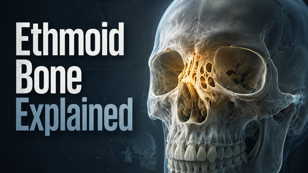
The ethmoid bone is a small, light, complex bone located between the eyes. It forms part of the roof of the nasal cavity, the medial walls of the orbits, the upper nasal septum, and the anterior skull base. Its close relationship with the olfactory nerves, ethmoid sinuses, eyes, and brain makes it especially important in anatomy, radiology, trauma assessment, and endoscopic sinus surgery.elsevier+1
Ethmoid Bone Basics
The ethmoid lies in the midline of the skull, behind the bridge of the nose and between the orbits. It sits below the frontal bone, in front of the sphenoid bone, above the nasal cavity, and beneath the anterior cranial fossa.
It contributes to several structures:
- Roof of the nasal cavity
- Upper bony part of the nasal septum
- Medial wall of each orbit
- Anterior cranial base
- Ethmoidal paranasal sinuses
- Passage of olfactory nerve fibres
The bone articulates with the frontal bone, sphenoid bone, vomer, nasal bones, lacrimal bones, maxillae, palatine bones, and inferior nasal conchae.
Why is it important?
This bone forms a central anatomical bridge between the nose, paranasal sinuses, orbit, and anterior skull base. Its thin bony walls and proximity to the olfactory nerves, eyes, and brain explain why disease, trauma, and surgery in this region require careful assessment.
Read more: Ethmoid Bone
Ethmoid Bone Location
The ethmoid bone is located:
- Behind the upper nose
- Between the eye sockets
- Below the frontal bone
- In front of the sphenoid bone
- Above the nasal cavity
- At the front of the skull base
It is best understood as the bone that connects the nasal cavity with the orbit and anterior cranial fossa.
Internal-link opportunity: link “anterior cranial fossa,” “orbit anatomy,” “nasal septum,” and “paranasal sinuses” to relevant supporting pages.
Parts of the Ethmoid Bone
The bone has four principal components:
- Cribriform plate
- Crista galli
- Perpendicular plate
- Two ethmoidal labyrinths
The ethmoidal labyrinths contain ethmoid air cells and contribute to the superior and middle nasal conchae, as well as the lamina papyracea.
Cribriform Plate Anatomy
The cribriform plate is a thin, horizontal plate that forms part of the roof of the nasal cavity and the floor of the anterior cranial fossa. Its sieve-like openings, called cribriform foramina, transmit small olfactory nerve fibres from the upper nasal cavity to the olfactory bulbs.
Functions of the cribriform plate
- Forms part of the nasal cavity roof
- Separates the nasal cavity from the anterior cranial fossa
- Transmits fibres of the olfactory nerve, cranial nerve I
- Supports the olfactory bulbs above it
Clinical importance
A cribriform plate fracture may disrupt olfactory nerve fibres, causing reduced smell or loss of smell. It can also create an abnormal connection between the nasal cavity and intracranial space, which may result in cerebrospinal fluid rhinorrhoea.
Clear, persistent watery nasal drainage after a significant head injury should be assessed urgently, particularly if it is associated with loss of smell, headache, fever, neck stiffness, or confusion.
Crista Galli
The crista galli is a vertical midline projection extending upward from the cribriform plate. Its name means “crest of the rooster.”
The falx cerebri, a dural fold that separates the right and left cerebral hemispheres, attaches to the crista galli.
Functions of the crista galli
- Provides attachment for the falx cerebri
- Acts as a midline landmark in the anterior cranial fossa
- Helps orient radiological and surgical assessment of the anterior skull base
Perpendicular Plate
The perpendicular plate is a thin vertical plate extending downward from the cribriform plate. It forms the upper part of the bony nasal septum and joins the vomer and septal cartilage to help divide the nasal cavity into right and left passages.elsevier+1
Functions of the perpendicular plate
- Forms part of the bony nasal septum
- Helps separate the right and left nasal cavities
- Supports normal nasal airflow
- Connects the upper skull base with the septal structures below
Clinical importance
Trauma or developmental variation involving this plate may contribute to septal deviation. A clinically significant deviated septum may be associated with one-sided nasal obstruction, altered airflow, recurrent nosebleeds, snoring, or difficulty with sinus drainage.
Ethmoidal Labyrinths
The ethmoidal labyrinths are paired, irregular masses on either side of the perpendicular plate. They contain multiple small air-filled cavities called ethmoid air cells, which make up the ethmoid sinuses.
The labyrinths contribute to:
- The ethmoid sinus system
- The lateral wall of the nasal cavity
- The medial orbital wall
- Superior nasal concha
- Middle nasal concha
Ethmoid Sinus Anatomy
Ethmoid air cells are commonly grouped as anterior, middle, and posterior cells.
| Group | Typical drainage pathway |
| Anterior ethmoid air cells | Middle meatus, commonly through the ethmoidal infundibulum |
| Middle ethmoid air cells | Middle meatus, often around the ethmoidal bulla |
| Posterior ethmoid air cells | Superior meatus |
Blocked drainage pathways can contribute to inflammation or infection of the ethmoid sinuses.
Lamina Papyracea Anatomy
The lamina papyracea is the delicate lateral wall of the ethmoidal labyrinth. It forms much of the medial wall of the orbit and separates the ethmoid sinuses from the eye socket.
Its name means “paper plate,” reflecting its thin, fragile structure.
Why does the lamina papyracea matter?
Because this bony plate is thin, severe ethmoid sinus infection can extend toward the orbit. Eye symptoms occurring with sinus disease require prompt medical assessment.
Potential warning signs include:
- Eyelid swelling or redness
- Eye pain
- Pain during eye movement
- Double vision
- Reduced vision
- Reduced eye movement
- Fever or worsening systemic illness
Surgical relevance
The lamina papyracea is a key landmark in endoscopic sinus surgery. Injury may expose orbital fat or, less commonly, affect extraocular muscles or vision. Preoperative CT assessment helps surgeons identify the relationship between the ethmoid sinuses, orbit, and skull base.
Internal-link opportunity: link “orbital cellulitis,” “sinus infection,” and “endoscopic sinus surgery” to related clinical pages.
Superior and Middle Nasal Conchae
The superior and middle nasal conchae are curved bony shelves formed by the ethmoidal labyrinths. They project from the lateral wall of the nasal cavity.
The inferior nasal concha is a separate bone and is not part of the ethmoid bone.
Functions of the nasal conchae
The conchae increase the surface area within the nasal cavity and help to:
- Warm inhaled air
- Humidify inhaled air
- Filter particles
- Direct airflow
- Support normal sinus ventilation and drainage
The middle turbinate is particularly important in nasal endoscopy because it is a major landmark for the middle meatus and nearby sinus drainage pathways.
Ethmoid Bone Articulations
| Bone | Relationship |
| Frontal bone | Articulates superiorly at the anterior skull base |
| Sphenoid bone | Articulates posteriorly |
| Vomer | Contributes to the bony nasal septum |
| Nasal bones | Articulates anteriorly |
| Lacrimal bones | Contributes to the medial orbital region |
| Maxillae | Articulates at orbital and nasal surfaces |
| Palatine bones | Connects posteriorly within the nasal cavity |
| Inferior nasal conchae | Articulates along the lateral nasal wall |
These extensive articulations explain the bone’s central role in facial, nasal, orbital, and skull-base anatomy.
Ethmoid Bone Function
The ethmoid has several important functions.
Supports the sense of smell
Olfactory nerve fibres pass through the cribriform plate to reach the olfactory bulbs. This pathway allows smell information from the nasal cavity to reach the brain.
Forms part of the nasal cavity
The perpendicular plate contributes to the nasal septum, while the labyrinths and conchae contribute to the lateral nasal walls.
Contributes to the medial orbital wall
The lamina papyracea forms a major portion of the medial orbital wall and separates the orbit from the ethmoid sinuses.
Forms part of the anterior skull base
The cribriform plate separates the nasal cavity from the anterior cranial fossa and contributes to the skull base beneath the frontal lobes.
Contains ethmoid air cells
The ethmoidal labyrinths contain the ethmoid air cells, a component of the paranasal sinus system.
Helps condition inhaled air
The superior and middle nasal conchae increase mucosal surface area, helping warm, humidify, and filter inhaled air.
Development
The ethmoid develops from cartilaginous elements of the embryonic nasal capsule. Ossification begins during fetal life, while the ethmoid air-cell system develops further after birth and continues to enlarge through childhood.
This is clinically relevant in paediatric imaging because sinus anatomy and aeration differ by age. Imaging findings should therefore be interpreted in the context of normal developmental anatomy.
Clinical Importance
The ethmoid region is important in sinus disease, trauma, imaging, and surgery because it lies next to the orbit, skull base, olfactory nerves, and nasal cavity.
Ethmoid Sinusitis
Ethmoid sinusitis refers to inflammation or infection of the ethmoid air cells. It may occur with viral upper-respiratory infections, allergic inflammation, bacterial rhinosinusitis, nasal polyps, or impaired sinus drainage.
Symptoms can include:
- Nasal blockage
- Pressure between or behind the eyes
- Reduced smell
- Headache
- Thick nasal discharge
- Postnasal drip
- Fever in some infections
Orbital complications
Ethmoid sinus disease can occasionally spread to tissues around the eye. Seek urgent assessment for eye swelling, redness, pain with eye movement, double vision, visual change, impaired eye movement, severe headache, high fever, or rapid clinical deterioration.
Cribriform plate injury
Head trauma affecting the anterior skull base may injure the cribriform plate. Possible features include loss of smell or clear nasal drainage that could represent a cerebrospinal fluid leak.
Urgent assessment is required after significant head trauma, especially when neurological symptoms, persistent fluid leakage, fever, neck stiffness, or altered consciousness are present.
Anosmia and olfactory dysfunction
Anosmia is loss of smell. Problems in the ethmoid region may affect smell because the olfactory nerve fibres traverse the cribriform plate.
Causes may include:
- Head trauma
- Chronic rhinosinusitis
- Nasal polyps
- Viral illness
- Tumours
- Surgery involving the nasal cavity or skull base
Deviated nasal septum
Because the perpendicular plate contributes to the bony nasal septum, developmental variation or injury can affect septal alignment. A deviated septum may contribute to nasal obstruction, recurrent epistaxis, snoring, or impaired airflow.
Tumours of the ethmoid region
Benign and malignant lesions can affect the ethmoid region. Persistent one-sided nasal obstruction, repeated unexplained nosebleeds, facial swelling, reduced smell, persistent pain, or eye symptoms should be evaluated clinically.
Imaging and Radiology
CT is especially useful for assessing bony anatomy, sinus opacification, fractures, anatomical variants, and preoperative planning. MRI provides better soft-tissue detail when assessing the orbit, brain, nerves, inflammatory changes, tumours, or suspected intracranial extension.
| Method | Main role |
| CT | Bony anatomy, fractures, sinus disease, air cells, skull-base landmarks, surgical planning |
| MRI | Soft tissues, orbit, brain, nerves, tumours, inflammation, possible intracranial extension |
| Nasal endoscopy | Direct assessment of nasal passages, middle meatus, polyps, inflammation, and accessible lesions |
Important CT landmarks
Radiologists and surgeons commonly assess:
- Cribriform plate
- Crista galli
- Lamina papyracea
- Ethmoid air cells
- Middle turbinate
- Uncinate process
- Ethmoidal infundibulum
- Ethmoidal bulla
- Orbit
- Ethmoid roof
- Lateral lamella
Keros Classification
The Keros classification describes olfactory fossa depth, which is determined by the height of the lateral lamella of the cribriform plate. It is relevant to preoperative CT review because the lateral lamella is among the thinnest parts of the anterior skull base.pmc.ncbi.nlm.nih+1
| Keros type | Olfactory fossa depth | Clinical relevance |
| Type I | 1–3 mm | Shallower olfactory fossa |
| Type II | 4–7 mm | Intermediate depth |
| Type III | 8–16 mm | Deeper fossa and a longer lateral lamella |
A deeper olfactory fossa may increase the need for surgical caution because an extended, thin lateral lamella is more vulnerable to skull-base injury during endoscopic ethmoid surgery.pmc.ncbi.nlm.nih+1
Ethmoid Bone and Sinus Surgery
The ethmoid region is central to functional endoscopic sinus surgery. Procedures may be used in selected patients with chronic rhinosinusitis, nasal polyps, recurrent sinus disease, fungal sinus disease, selected mucoceles, or skull-base pathology.
Important surgical landmarks
| Structure | Surgical importance |
| Middle turbinate | Main orientation landmark during endoscopy |
| Uncinate process | Helps define the anterior drainage pathway |
| Ethmoidal bulla | Prominent middle-ethmoid landmark |
| Lamina papyracea | Identifies the medial orbital boundary |
| Cribriform plate | Identifies the anterior skull base |
| Lateral lamella | Thin skull-base region vulnerable to injury |
| Basal lamella of middle turbinate | Helps separate anterior and posterior ethmoid regions |
Potential surgical risks
Surgery in this area is performed near the orbit and skull base. Potential complications include bleeding, orbital injury, cerebrospinal fluid leak, infection, smell disturbance, scar formation, persistent symptoms, eye movement problems, or visual complications.
Individual risk depends on disease severity, anatomical variation, imaging findings, surgical extent, and the patient’s overall health.
Ethmoid Bone Mnemonic
A concise way to remember the main components is:
C-P-L: Cribriform plate, Perpendicular plate, Labyrinths
A functional memory aid is:
Cribriform smells, Perpendicular separates, Labyrinth aerates.
- The cribriform plate carries olfactory nerve fibres.
- The perpendicular plate contributes to the nasal septum.
- The ethmoidal labyrinths contain ethmoid air cells.
Another useful reminder is:
“Paper wall beside the eye” = lamina papyracea.
Clinical Anatomy Summary
| Structure | Primary role | Clinical importance |
| Cribriform plate | Roof of the nasal cavity; passage for olfactory nerve fibres | Trauma may cause smell loss or CSF leak |
| Crista galli | Attachment for falx cerebri | Midline skull-base landmark |
| Perpendicular plate | Upper bony nasal septum | May contribute to septal deviation |
| Lamina papyracea | Medial orbital wall | Thin barrier between orbit and ethmoid sinuses |
| Lateral lamella | Part of the olfactory fossa boundary | Important skull-base risk area in surgery |
| Ethmoid air cells | Paranasal sinus spaces | Can be affected by sinus inflammation or infection |
| Middle turbinate | Lateral nasal-wall structure | Key endoscopic surgical landmark |
Ethmoid Bone Exam Review
| Question | Answer |
| Question | Answer |
| Where is the ethmoid bone located? | Between the orbits, above the nasal cavity, and below the anterior cranial fossa |
| Which cranial nerve passes through the cribriform plate? | The olfactory nerve, CN I |
| What attaches to the crista galli? | The falx cerebri |
| What forms much of the medial orbital wall? | The lamina papyracea |
| Which component helps form the nasal septum? | The perpendicular plate |
| Which structures contain the ethmoid air cells? | The paired ethmoidal labyrinths |
| Which nasal conchae are ethmoidal structures? | The superior and middle nasal conchae |
| Is the inferior nasal concha part of this bone? | No; it is a separate facial bone |
| What assesses olfactory fossa depth? | Keros classification |
| What may follow cribriform plate trauma? | Olfactory dysfunction and possible CSF rhinorrhoea |
Frequently Asked Questions
What is the ethmoid bone?
The ethmoid bone is a central skull bone located between the eyes. It helps form the nasal cavity, upper nasal septum, medial orbital walls, ethmoid sinuses, and part of the anterior skull base. Its cribriform plate also provides a route for olfactory nerve fibres, making it essential to the sense of smell.
What are the main parts of the ethmoid bone?
Its principal components are the cribriform plate, crista galli, perpendicular plate, and paired ethmoidal labyrinths. The labyrinths contain ethmoid air cells, contribute to the lateral nasal walls, and include the lamina papyracea, which forms much of the medial wall of the orbit.
What passes through the cribriform plate?
Fine filaments of the olfactory nerve, also called cranial nerve I, pass through the cribriform foramina. These fibres carry smell signals from receptors in the upper nasal cavity to the olfactory bulbs. Injury to this area can impair or eliminate the sense of smell.
What is the lamina papyracea?
The lamina papyracea is a very thin bony plate of the ethmoidal labyrinth. It forms a major part of the medial orbital wall and separates the orbit from the ethmoid sinuses. Its thinness is important in severe sinus infection and during endoscopic sinus surgery.
Can ethmoid sinusitis affect the eyes?
Yes. Severe infection in the ethmoid air cells can extend toward the orbit because the lamina papyracea is thin. Urgent clinical assessment is needed if sinus symptoms occur with eye swelling, redness, pain on eye movement, double vision, difficulty moving the eye, reduced vision, high fever, or worsening illness.
Can a cribriform plate fracture cause a CSF leak?
Yes. A fracture may create a connection between the intracranial space and nasal cavity, allowing cerebrospinal fluid to drain through the nose. Persistent clear, watery nasal drainage after head trauma should be assessed urgently, particularly if it occurs with loss of smell, headache, fever, neck stiffness, or neurological symptoms.
Which nasal conchae are formed by the ethmoid bone?
The superior and middle nasal conchae are parts of the ethmoid bone. The inferior nasal concha is not; it is an independent bone attached to the lateral wall of the nasal cavity. The conchae help warm, humidify, filter, and direct inspired air.
What does Keros classification measure?
Keros classification groups the depth of the olfactory fossa according to the height of the lateral lamella of the cribriform plate. It is used in radiological assessment before endoscopic sinus surgery because a deeper fossa may indicate a longer, more vulnerable lateral lamella.pmc.ncbi.nlm.
How is the ethmoid bone seen on imaging?
CT is preferred for assessing the bone’s fine bony anatomy, ethmoid air cells, sinus disease, fractures, and skull-base landmarks. MRI is more useful when clinicians need to assess soft tissues, including the orbit, olfactory region, brain, inflammation, tumours, or suspected intracranial extension.
Why is the ethmoid bone important in sinus surgery?
It contains the ethmoid air cells and lies beside the orbit and skull base. Surgeons use landmarks such as the middle turbinate, uncinate process, ethmoidal bulla, lamina papyracea, cribriform plate, and lateral lamella to navigate safely during endoscopic sinus surgery.