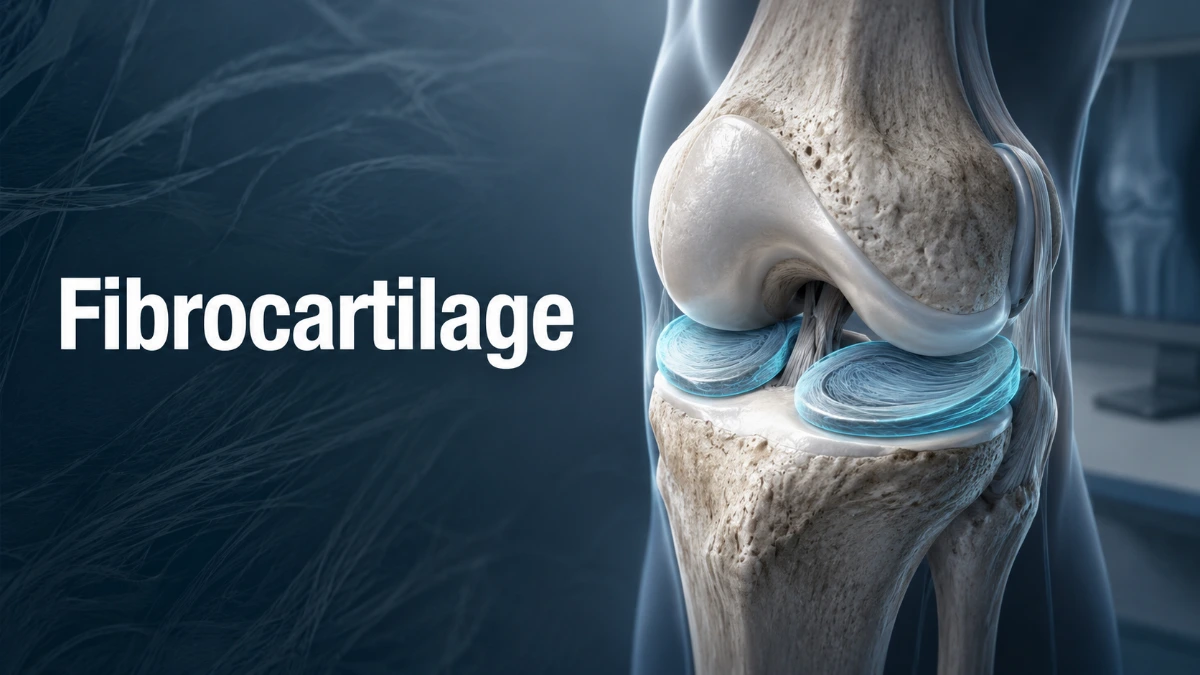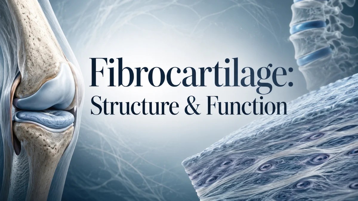
Fibrocartilage is a specialized type of cartilage that helps the body tolerate high compression, tension, and shear forces. It is found in the knee menisci, intervertebral discs, pubic symphysis, joint labra, and the triangular fibrocartilage complex (TFCC) of the wrist. Its dense collagen-rich matrix makes it highly durable, but its limited blood supply also contributes to slow healing after injury.
What Is Fibrocartilage?
Fibrocartilage is a strong supportive connective tissue with features of both dense fibrous connective tissue and cartilage. It contains chondrocytes—cartilage cells—within small spaces called lacunae. These cells maintain an extracellular matrix dominated by thick collagen bundles, especially type I collagen.
Its structure makes it well suited for anatomical sites exposed to repeated loading. In the knee, it helps distribute body weight. In the spine, it reinforces the outer portion of the intervertebral disc. In the wrist, it helps stabilize the ulnar side during gripping and forearm rotation.
Rather than acting as a smooth gliding surface like articular cartilage, this tissue works as a load-management system. It can deform slightly under pressure, disperse force across a wider area, and resist splitting or tearing under repeated stress.
Fibrocartilage Histology at a Glance
Fibrocartilage has a distinct microscopic appearance. Chondrocytes are commonly arranged in rows or small groups between dense bundles of collagen fibers, giving the tissue a more fibrous and less glassy appearance than hyaline cartilage.
| Histological feature | Significance |
| Chondrocytes in lacunae | Maintain the extracellular matrix |
| Predominantly type I collagen | Provides high tensile strength |
| Variable type II collagen | Supports cartilage-like matrix properties in some regions |
| Sparse ground substance | Produces a denser, more fibrous texture than hyaline cartilage |
| Proteoglycans and water | Help the tissue resist compression |
| Rows of chondrocytes | Often follow the orientation of collagen fibers and local loading patterns |
| No typical perichondrium | Limits access to cells that could support repair |
The extracellular matrix contains collagen, water, proteoglycans, and glycosaminoglycans. Proteoglycans, including aggrecan in many cartilaginous tissues, attract water and contribute to resistance against compression. However, compared with hyaline cartilage, fibrocartilage contains more dense collagen and less proteoglycan-rich ground substance.
- Read more: Fibrocartilage
Why Is Fibrocartilage White?
This tissue appears white and opaque because of its densely packed collagen fibers. The fibers scatter light more strongly than the smoother matrix of hyaline cartilage, which has a more glassy and translucent appearance.
The white appearance reflects its mechanical role. Dense collagen provides resilience against pulling, twisting, compression, and shear forces.
Fibrocartilage Function
The main function of fibrocartilage is to distribute mechanical load while resisting compression, tension, and shear. It helps stabilize joints, absorb shock, protect adjacent bone and articular cartilage, and reduce stress concentration where forces are repeatedly applied.
Compression, Tension, and Shear Resistance
Fibrocartilage is especially valuable where the body must manage multiple types of force at the same time.
- Compression occurs when force presses tissues together, such as body weight acting through the knee or spine.
- Tension occurs when a structure is pulled, such as a tendon or ligament transmitting force to bone.
- Shear occurs when nearby surfaces move in opposing directions, as during knee rotation or wrist motion.
- Torsion occurs when a structure is twisted, as during spinal rotation or forearm pronation and supination.
Its collagen fibers often follow local force directions. This gives the tissue directional mechanical behavior: it may resist force differently depending on the angle and direction of loading.
For example, the menisci use their collagen arrangement to manage compression and convert part of that load into circumferential tensile stress. In the annulus fibrosus, collagen layers arranged in alternating directions help resist twisting and bending forces in the spine.
Why Fibrocartilage Exists Instead of Bone or Hyaline Cartilage
Bone is highly resistant to compression but relatively rigid. If bone occupied every high-load interface, force would be transmitted more directly and joint movement would become less forgiving.
Hyaline cartilage creates smooth, low-friction gliding surfaces, but it is not designed to withstand the same combination of high tensile, compressive, and shear forces seen in the menisci, labra, and intervertebral discs.
Fibrocartilage provides a practical middle ground. It can deform enough to spread load and improve joint congruence, while its collagen-rich matrix helps prevent excessive deformation and tearing.
The knee illustrates this role clearly. The femur and tibia do not have perfectly matching surfaces. Meniscal tissue improves their fit, spreads body weight over a broader contact area, and helps protect the joint from concentrated stress.
Fibrocartilage Locations in the Body
Fibrocartilage is concentrated in areas that need durable cushioning, reinforcement, or a gradual transition between flexible and rigid tissue.
| Location | Main role |
| Intervertebral discs | The annulus fibrosus resists tension and helps contain the nucleus pulposus |
| Knee menisci | Distribute load, improve joint congruence, support stability, and absorb shock |
| Pubic symphysis | Connects the pubic bones while allowing limited pelvic movement |
| Glenoid labrum of the shoulder | Deepens the shoulder socket and contributes to stability |
| Acetabular labrum of the hip | Deepens the hip socket and supports load distribution |
| Triangular fibrocartilage complex of the wrist | Supports the distal radioulnar joint and ulnar side of the wrist |
| Fibrocartilaginous entheses | Help transfer force where tendons and ligaments attach to bone |
| Selected articular discs | Improve load distribution and congruence in specific synovial joints |
Fibrocartilaginous Entheses
An enthesis is where a tendon or ligament attaches to bone. At some high-load attachment sites, fibrocartilage forms a gradual transition between flexible connective tissue and rigid bone.
This transition helps prevent stress from concentrating in one small area. Instead of force moving abruptly from a flexible tendon into stiff bone, the tissue changes progressively through zones with different compositions and mechanical properties.
Sharpey’s fibers help anchor collagen-rich structures into bone, while fibrocartilage helps manage compression and repeated loading at the attachment site.
Fibrocartilage vs Hyaline vs Elastic Cartilage
The body has three major cartilage types: fibrocartilage, hyaline cartilage, and elastic cartilage. All contain chondrocytes and extracellular matrix, but each is adapted for different mechanical demands.
| Feature | Fibrocartilage | Hyaline cartilage | Elastic cartilage |
| Main fibers | Predominantly type I collagen, with variable type II collagen | Mainly type II collagen | Type II collagen with elastic fibers |
| Appearance | White, dense, fibrous, opaque | Glassy, smooth, translucent | Flexible and elastic |
| Matrix | Relatively sparse ground substance | More abundant proteoglycan-rich ground substance | Elastic-fiber network with cartilage matrix |
| Tensile strength | Very high | Moderate | Moderate |
| Compression resistance | High | High | Moderate |
| Flexibility | Limited to moderate | Moderate | High |
| Perichondrium | Usually absent | Usually present, except at articular surfaces | Present |
| Blood supply | Avascular | Avascular | Avascular |
| Typical locations | Menisci, discs, pubic symphysis, labra, TFCC | Articular surfaces, costal cartilage, trachea, nose | External ear, epiglottis |
| Main purpose | Load distribution and resistance to mixed forces | Smooth joint motion and structural support | Flexible support and shape retention |
| Healing capacity | Limited | Limited | Limited |
Cartilage does not have a direct blood supply. It receives nutrients and removes waste mainly through diffusion from adjacent tissues or fluids. This supports normal function but limits repair after substantial injury.
Fibrocartilage vs Dense Connective Tissue
Fibrocartilage and dense connective tissue both contain abundant collagen, but they differ in cellular organization and matrix composition.
Fibrocartilage contains chondrocytes within lacunae and a cartilage-like extracellular matrix. Dense connective tissue contains fibroblasts arranged among collagen fibers without the characteristic lacunae of cartilage.
A tendon is an example of dense regular connective tissue. It is optimized to transmit tension in a predictable direction. Fibrocartilage is more suitable where compression, tension, and shear occur together, such as where a tendon bends around bone or transitions into a bony attachment.
How Fibrocartilage Develops
Fibrocartilage develops from mesenchymal precursor cells that differentiate under the influence of local biological signals and mechanical conditions. Its formation is closely related to the loads a tissue must manage over time.
At tendon and ligament attachments, recurrent compression against bone can promote fibrocartilaginous adaptation. In the menisci, labra, discs, and articular structures, developmental pathways establish collagen orientation and matrix composition that match long-term mechanical demands.
Cells respond to physical forces through mechanotransduction. This process allows mechanical loading to influence cellular activity, matrix production, tissue maintenance, and adaptation. Normal loading helps maintain tissue organization, while excessive, repetitive, or abnormal loading can contribute to structural degeneration.
Blood Supply, Nutrition, and Healing Capacity
Fibrocartilage heals poorly for several connected reasons. Limited blood supply is important, but it is not the only explanation.
- It is largely avascular, so relatively few blood-borne repair cells and signaling molecules reach an injury.
- Nutrients move mainly by diffusion, which is slower than direct circulation.
- It has low cellularity compared with highly vascular tissues.
- Chondrocytes have limited ability to rapidly rebuild large amounts of matrix.
- It generally lacks a perichondrium, which can provide progenitor cells in some other cartilage types.
- Its extracellular matrix has slow turnover, particularly in mature load-bearing tissue.
- Ongoing loading can disrupt repair if a tear occurs in an unstable or poorly vascularized region.
Healing potential also varies within the same structure. The outer portion of the knee meniscus has a better blood supply than its central region, so peripheral tears may heal more successfully than tears in the inner avascular zone.
Does Fibrocartilage Contain Nerves?
The central fibrocartilage matrix is generally not richly innervated. However, surrounding tissues—including the capsule, synovium, ligaments, bone, attachments, and more vascular peripheral regions—contain nerve endings.
This is why injury can still be painful. Pain may arise from inflamed tissue at the margins of a tear, altered joint mechanics, instability, irritation of nearby nerves, or loading of adjacent structures.
Aging and Degeneration
Fibrocartilage can change with age, repeated loading, injury, altered alignment, and some systemic disorders. These changes may involve collagen disorganization, reduced water content, altered proteoglycan composition, slower cell activity, matrix breakdown, and, in some sites, calcification.
Degenerative changes do not always cause symptoms. Meniscal or disc abnormalities may appear on imaging in people with little or no pain. For this reason, clinicians interpret imaging alongside symptoms, movement limitations, medical history, and physical examination findings.
Aging does not mean fibrocartilage will inevitably fail. However, reduced tissue resilience and accumulated mechanical stress may make some structures more vulnerable to injury or degeneration.
Common Fibrocartilage Conditions
Fibrocartilage conditions can occur after acute trauma, repetitive loading, altered biomechanics, age-related change, or a combination of these factors.
Meniscal Tears
A meniscal tear is one of the most common fibrocartilage injuries. It may occur during twisting, pivoting, squatting, or contact injury. Degenerative tears can also develop gradually, especially when tissue resilience declines over time.
Possible symptoms include knee pain, swelling, stiffness, catching, locking, reduced motion, and a sensation that the knee may give way. The pattern and location of the tear, knee stability, symptoms, activity needs, and the condition of surrounding cartilage all influence management.
Treatment may include activity modification, rehabilitation, medication for symptom relief when appropriate, or surgery in selected cases. When surgery is needed, preserving as much functional meniscal tissue as possible is generally preferred.
Annular Tears, Disc Degeneration, and Disc Herniation
The outer portion of an intervertebral disc, called the annulus fibrosus, contains fibrocartilage. Repetitive loading, trauma, aging, and altered spinal mechanics can contribute to annular fissures or tears.
Disc degeneration may involve changes in hydration, collagen organization, matrix composition, and force distribution. A disc herniation occurs when disc material extends beyond its normal boundary, often through a weakened region of the annulus.
Symptoms vary by spinal level and whether nearby nerves are irritated or compressed. Back or neck pain may occur with or without radiating limb pain, numbness, tingling, or weakness. Imaging findings should always be interpreted in the context of symptoms and neurological examination.
Triangular Fibrocartilage Complex Tears
The triangular fibrocartilage complex (TFCC) is a fibrocartilaginous and ligamentous structure on the ulnar, or little-finger, side of the wrist. It helps stabilize the distal radioulnar joint, supports load transfer across the wrist, and contributes to smooth forearm rotation.
A TFCC tear may follow a fall onto an outstretched hand, forceful wrist rotation, repetitive gripping, or gradual degeneration. Common symptoms include ulnar-sided wrist pain, clicking, pain with gripping, and discomfort during pronation or supination.
The central part of the TFCC has limited blood supply, while peripheral regions are relatively more vascular. This difference can affect healing potential and influence treatment decisions.
Hip Labral Tears
The acetabular labrum is a fibrocartilaginous rim around the hip socket. A tear may be associated with trauma, repetitive hip movement, femoroacetabular impingement, hip dysplasia, or degenerative change.
Possible symptoms include groin pain, stiffness, clicking, catching, and discomfort during hip flexion or rotation. Similar symptoms can arise from several hip conditions, so a clinical assessment is needed before attributing pain to a labral tear alone.
Shoulder Labral Tears
The glenoid labrum is a fibrocartilaginous rim that deepens the shallow shoulder socket and supports stability. Tears may result from shoulder dislocation, trauma, repetitive overhead activity, or age-related change.
Symptoms may include deep shoulder pain, catching, clicking, weakness, instability, or discomfort with overhead movement. The type and location of a tear influence evaluation and treatment.
Pubic Symphysis Dysfunction
The pubic symphysis is a fibrocartilaginous joint that supports pelvic stability while permitting slight movement. Pain or dysfunction in this area may be associated with pregnancy-related pelvic changes, overuse in sports, trauma, or altered pelvic mechanics.
Symptoms can include pain at the front of the pelvis, discomfort when walking, turning in bed, climbing stairs, or standing on one leg. Persistent pelvic pain should be assessed because several conditions can cause similar symptoms.
Imaging and Diagnosis
Imaging can help assess fibrocartilage-related injuries, but it does not replace a clinical examination. A diagnosis should integrate symptoms, medical history, movement assessment, physical examination, and imaging when appropriate.
| Structure | Common assessment methods | Important consideration |
| Knee meniscus | Physical examination, MRI, and arthroscopy in selected cases | Imaging should be interpreted with symptoms |
| Intervertebral disc | Neurological examination and MRI when clinically appropriate | Disc changes can occur without pain |
| TFCC | Wrist examination, radiographs, MRI, MR arthrography, or arthroscopy in selected cases | Small or complex tears may be difficult to define on standard MRI |
| Hip labrum | Clinical examination, radiographs, MRI, or MR arthrography | Other hip conditions can produce similar symptoms |
| Shoulder labrum | Clinical examination, radiographs, MRI, or MR arthrography | Imaging should be correlated with instability and function |
| Pubic symphysis | Clinical examination and imaging in selected cases | Pelvic pain can have multiple causes |
For persistent symptoms, the most useful diagnostic approach depends on the involved body region and suspected condition. Imaging should answer a clinical question rather than serve as a stand-alone explanation for pain.
Treatment Principles
Treatment depends on the location of the problem, type of injury, severity of symptoms, joint stability, activity goals, tissue quality, and associated conditions. A scan alone does not determine the best treatment.
Conservative care may include:
- Temporary activity modification
- Guided rehabilitation and progressive strengthening
- Restoration of mobility and movement control
- Load-management strategies
- Bracing or splinting for selected injuries
- Clinician-recommended pain management
Surgery may be considered when symptoms remain substantial despite appropriate nonoperative care, when there is mechanical locking or instability, or when a tear has a repairable pattern with meaningful healing potential.
Can Exercise Repair Fibrocartilage?
Exercise cannot reliably restore a torn meniscus, TFCC, labrum, or degenerated disc to its original structure. However, properly prescribed rehabilitation can improve strength, mobility, movement control, joint function, and load tolerance.
For example, hip and quadriceps strengthening may improve knee control in some people with meniscal symptoms. Wrist rehabilitation may help restore function after selected TFCC injuries. The correct programme depends on the diagnosis, stage of recovery, and symptoms; exercises introduced too early or performed incorrectly may aggravate some injuries.
Can Food or Supplements Repair Fibrocartilage?
No food or supplement has been proven to regrow damaged meniscal tissue, repair a TFCC tear, heal a labral tear, or restore a degenerated disc to its original state.
A balanced diet with adequate protein, energy, vitamins, and minerals supports general health and recovery. However, nutrition should not be presented as a replacement for medical assessment, rehabilitation, or treatment when symptoms are persistent or severe.
Can Fibrocartilage Regrow?
Fibrocartilage has limited regenerative capacity. Small injuries in relatively vascular peripheral areas may have better healing potential than injuries in central avascular regions. Even when healing occurs, the repaired tissue may not fully reproduce the original structure or mechanical behavior.
Research into cell-based therapies, biological scaffolds, growth factors, and other regenerative approaches is ongoing. These treatments are not universal solutions, and their evidence, availability, and suitability vary by condition and clinical setting.
When to Seek Medical Assessment
Seek a medical assessment for persistent or worsening symptoms, especially when there is:
- Joint locking or inability to fully move a joint
- Major swelling, deformity, or inability to bear weight after an injury
- Wrist instability or ongoing ulnar-sided wrist pain after trauma
- Recurrent catching, giving way, or painful clicking in the knee, shoulder, or hip
- Persistent pelvic pain affecting walking, sleep, or daily activities
- New weakness, numbness, or changes in bowel or bladder control with back or neck pain
Urgent assessment is particularly important after significant trauma or when neurological symptoms accompany spinal pain.
Frequently Asked Questions
What is the main function of fibrocartilage?
Fibrocartilage distributes force and resists combined compression, tension, and shear. It is used where hyaline cartilage would not provide enough tensile strength and where bone would be too rigid. In the knee, it helps spread body weight across the tibia. In the spine, it reinforces the annulus fibrosus around the nucleus pulposus. In the wrist, the TFCC helps stabilize the distal radioulnar joint during rotation and gripping.
Is fibrocartilage stronger than hyaline cartilage?
Fibrocartilage is generally more resistant to tensile and shear forces because its matrix contains abundant type I collagen. Hyaline cartilage is better suited to smooth, low-friction movement at articular surfaces. Neither tissue is universally stronger; each is specialized for a different role. Fibrocartilage is ideal for menisci and intervertebral discs, whereas hyaline cartilage is best suited to the gliding surfaces of synovial joints.
Where is fibrocartilage found in the human body?
Important locations include the knee menisci, annulus fibrosus of intervertebral discs, pubic symphysis, shoulder glenoid labrum, hip acetabular labrum, selected articular discs, tendon and ligament attachment sites, and the TFCC of the wrist. These structures all need durable cushioning and resistance to complex loading. Composition can vary within one structure because the direction and intensity of force differ across regions.
Why does fibrocartilage heal poorly?
It heals slowly because it has little direct blood supply and relies mainly on diffusion for nutrient delivery. It also has low cellularity, slow matrix turnover, limited capacity to replace damaged collagen, and usually lacks a perichondrium that could contribute repair cells. Healing differs by location. A peripheral meniscal tear may have a better chance of healing than a central tear in the avascular inner region.
Does fibrocartilage contain blood vessels or nerves?
Most fibrocartilage is avascular, though outer regions, attachments, and nearby tissues can contain blood vessels. Nerve supply is limited within the central matrix, but surrounding tissues such as ligaments, synovium, capsule, bone, and vascularized edges can generate pain. This is why meniscal, disc, labral, or TFCC injuries may be painful despite limited innervation within the tissue itself.
Can MRI always detect a TFCC tear?
No. MRI is useful for assessing the wrist, but the TFCC is small and anatomically complex, so subtle tears can be difficult to identify. Accuracy depends on imaging quality, technique, tear type, and interpretation. In selected cases, MR arthrography or wrist arthroscopy may provide additional detail. Diagnosis should combine symptoms, physical examination findings, imaging, and response to initial management.
What is the difference between the meniscus and TFCC?
Both are fibrocartilage-containing structures that distribute load and support joint stability, but they are located in different joints. The menisci lie between the femur and tibia in the knee, where they improve load distribution and joint congruence. The TFCC lies on the ulnar side of the wrist and helps support the distal radioulnar and ulnocarpal regions. Unlike a meniscus, the TFCC includes important ligamentous components as well as fibrocartilage.
Can fibrocartilage become calcified?
Cartilage can calcify with aging, degeneration, injury, or certain disease processes. Calcification may alter mechanical behavior and can occur alongside changes in the extracellular matrix. Its significance depends on the structure involved, the extent of calcification, associated symptoms, and other health conditions. An imaging finding of calcification should be interpreted in the full clinical context rather than treated as a diagnosis by itself.
Can exercise or diet rebuild damaged fibrocartilage?
Exercise and diet cannot reliably regrow a torn meniscus, repair a TFCC or labral tear, or restore a degenerated disc to its original condition. Rehabilitation can improve strength, mobility, coordination, and load tolerance, while balanced nutrition supports overall health. The best plan depends on the injury. For example, a displaced tear causing joint locking may require a different approach from gradual pain without mechanical symptoms.
What are common symptoms of a fibrocartilage injury?
Symptoms depend on the affected structure. Meniscal injuries can cause knee pain, swelling, catching, locking, or giving way. TFCC injuries may cause pain on the little-finger side of the wrist, clicking, and pain with gripping or rotation. Disc problems can cause back or neck pain and sometimes radiating pain, numbness, tingling, or weakness. Hip and shoulder labral injuries may cause deep joint pain, stiffness, clicking, catching, or instability.