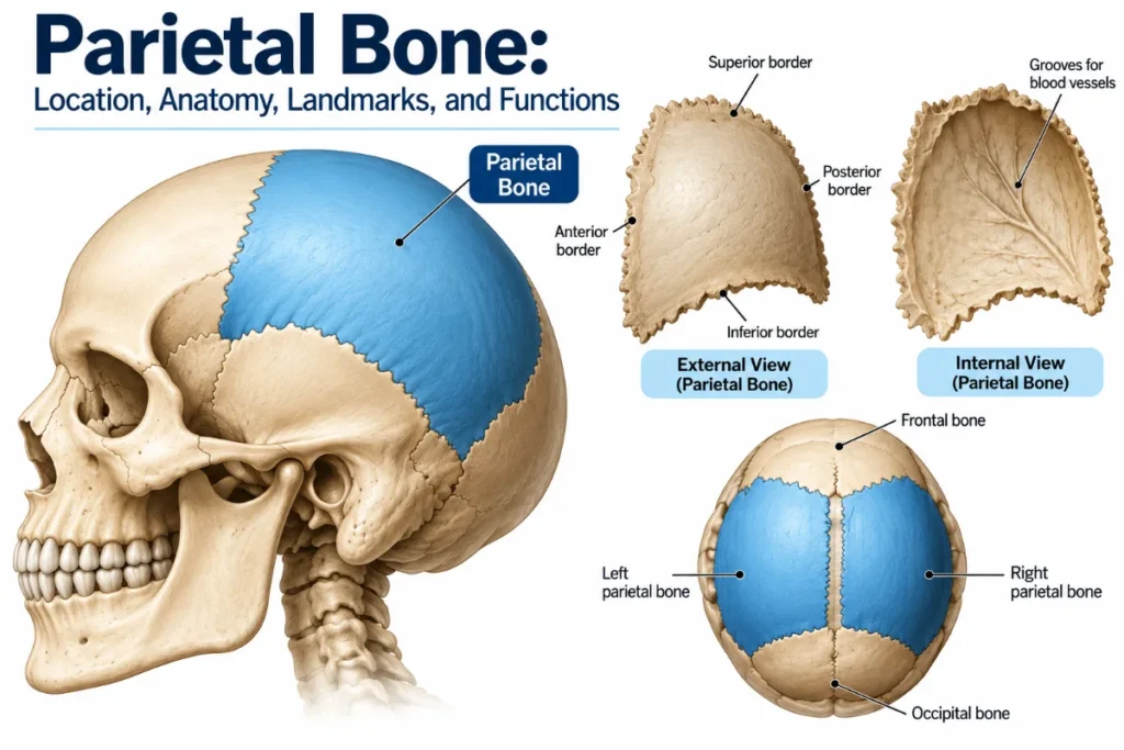
The parietal bone is one of two paired, curved flat bones that form much of the upper and lateral portions of the human skull. Located on either side of the head, these bones make up a large part of the cranial vault, the bony structure that surrounds and protects the brain. They also help maintain the skull’s shape and provide attachment areas for muscles and fascia.
The right and left parietal bones meet along the sagittal suture at the top of the skull. Each bone has two surfaces, four borders, four angles, and several recognizable landmarks. These features help explain how the bones fit together and support the surrounding structures.
Where Is the Parietal Bone Located?
The parietal bones form much of the roof and upper sides of the skull. Each extends from the frontal bone at the front of the head toward the occipital bone at the back. Below the parietal bone lies the temporal bone, which contributes to the lower side and base of the skull.
The main anatomical relationships include:
- Superiorly: The two parietal bones meet along the sagittal suture.
- Anteriorly: Each parietal bone connects with the frontal bone.
- Posteriorly: Both parietal bones meet the occipital bone.
- Inferiorly: Each articulates with the temporal and sphenoid bones.
Together, these bones form a substantial portion of the neurocranium, the part of the skull that encloses the brain. Although classified as flat bones, they curve to follow the rounded contour of the head. This arrangement is consistent with the anatomical description of the parietal bone.
Parietal Bone Anatomy and Major Landmarks
Each parietal bone is roughly quadrilateral, with two surfaces, four borders, and four angles. Its distinctive features make it possible to identify the bone and understand its connections with neighboring cranial structures.
External and Internal Surfaces
The parietal bone has a smooth, outward-facing convex surface and a concave internal surface that faces the brain.
External Surface
Several important landmarks appear on the outer surface:
- Parietal eminence: A rounded prominence near the center of the bone. It is a useful landmark and marks an early center of bone development.
- Superior temporal line: A curved ridge that provides an attachment area for the temporal fascia.
- Inferior temporal line: A curved ridge below the superior temporal line that marks part of the attachment of the temporalis muscle.
- Parietal foramen: A small opening that may occur near the posterior portion of the sagittal border. When present, it typically transmits an emissary vein.
The parietal foramen varies between individuals and may be absent. Its location and relationship with nearby veins have been documented in anatomical research on the parietal foramen.
Internal Surface
The inner surface is concave and contains grooves and depressions created by nearby blood vessels and structures associated with the brain’s protective membranes.
Its main features include:
- Branching grooves for the middle meningeal artery.
- A groove near the superior border for the superior sagittal sinus.
- A groove near the mastoid angle associated with the sigmoid sinus.
- Small depressions related to arachnoid granulations, usually near the sagittal border.
- Impressions corresponding to the underlying brain and nearby blood vessels.
The internal surface lies next to the dura mater, the tough outer membrane surrounding the brain. Its relationship with the middle meningeal artery and venous sinuses is especially important when assessing traumatic skull injuries.
Four Borders of the Parietal Bone
Each parietal bone has four borders that connect with neighboring cranial bones through fibrous joints called sutures.
| Border | Associated suture | Main connection |
| Sagittal (superior) | Sagittal suture | Opposite parietal bone |
| Frontal (anterior) | Coronal suture | Frontal bone |
| Occipital (posterior) | Lambdoid suture | Occipital bone |
| Squamous (inferior) | Squamous suture along its central portion | Temporal bone |
The inferior border also connects with the sphenoid bone anteriorly and the mastoid portion of the temporal bone posteriorly. These connections form the sphenoparietal and parietomastoid sutures, respectively.
These relationships are described in more detail in the anatomy of the parietal bone’s borders and sutures.
Four Angles of the Parietal Bone
The parietal bone has four angles, each marking a corner where two borders meet.
- Frontal angle: The upper front corner, near the bregma, where the sagittal and coronal sutures intersect.
- Sphenoidal angle: The lower front corner, contributing to the pterion region where several cranial bones meet.
- Occipital angle: The upper back corner, near the lambda, where the sagittal and lambdoid sutures meet.
- Mastoid angle: The lower back corner, contributing to the asterion region near the temporal and occipital bones.
These angles serve as useful reference points when identifying skull structures and the junctions between cranial bones.
Sutures and Connections With Other Skull Bones
Cranial sutures are strong fibrous joints that hold neighboring skull bones together. Four principal sutures help define the position of the parietal bones.
The sagittal suture connects the right and left parietal bones along the top of the skull. At the front, the coronal suture joins them to the frontal bone, while the lambdoid suture connects them with the occipital bone at the back.
The squamous suture runs along the lower side of the skull, connecting the parietal bone with the squamous portion of the temporal bone.
Together, these joints provide stability while allowing the skull to grow during childhood.
What Is the Function of the Parietal Bone?
The parietal bones have three main functions: protecting the brain, supporting skull structure, and providing attachment areas for surrounding soft tissues.
1. Protecting the brain
As part of the cranial vault, the parietal bones form a rigid protective covering over portions of the brain. Their curved structure helps shield delicate tissues from external forces, although a sufficiently strong impact can still cause injury.
2. Maintaining skull structure
The paired bones help give the skull its rounded shape and structural strength. Their connections with neighboring cranial bones create a stable framework that surrounds and protects the brain.
3. Providing muscle and fascia attachment areas
The temporal lines serve as attachment sites for structures associated with chewing. The superior temporal line provides an attachment for the temporal fascia, while the inferior temporal line marks part of the attachment of the temporalis muscle.
The temporalis muscle helps raise and retract the lower jaw, making it important for chewing.
Parietal Bone Fractures and Injuries
A parietal bone fracture occurs when one of the parietal bones breaks following head trauma. Common causes include falls, motor vehicle collisions, sports injuries, and direct blows to the head.
Symptoms may include pain, swelling, tenderness, bruising, or an open wound near the injured area. More severe trauma can also cause bleeding inside the skull or damage to the underlying brain.
A skull fracture does not always involve a brain injury. However, symptoms alone cannot reliably rule out serious complications, which is why suspected fractures require medical assessment.
How Are Parietal Bone Fractures Diagnosed?
Clinicians assess a suspected fracture by examining the head, reviewing how the injury occurred, and checking neurological function. They also look for symptoms that could suggest bleeding or damage inside the skull.
When imaging is necessary, a CT scan of the head without intravenous contrast is commonly used to identify skull fractures and assess possible intracranial injuries.
According to the clinical appropriateness criteria for head trauma imaging, noncontrast head CT is generally appropriate when clinical findings or established decision rules indicate the need for imaging.
Not every head injury requires a CT scan. The decision depends on the patient’s age, symptoms, examination findings, injury severity, and other clinical risk factors.
When Is a Head Injury an Emergency?
Seek immediate emergency medical care if any of these warning signs appear after a head injury:
- A worsening headache that does not go away.
- Repeated vomiting.
- Seizures or convulsions.
- Increasing confusion, unusual behavior, or slurred speech.
- Weakness, numbness, or difficulty coordinating movements.
- Unequal pupil sizes.
- Loss of consciousness, increasing drowsiness, or difficulty waking up.
These symptoms may indicate a serious head or brain injury and are not unique to parietal bone fractures. The emergency warning signs of traumatic brain injury also emphasize the need for urgent assessment when neurological symptoms develop or worsen.
Frequently Asked Questions
How many parietal bones are in the human skull?
The human skull normally contains two parietal bones, one on each side of the head. They meet at the sagittal suture and form much of the upper and lateral cranial vault.
Is the parietal bone a flat bone?
Yes. The parietal bone is classified as a flat bone, even though it has a curved shape. It consists of inner and outer layers of compact bone separated by a layer of spongy bone called diploë.
What is the difference between the parietal bone and the parietal lobe?
The parietal bone is part of the skull and helps protect the brain. The parietal lobe is a region of the brain involved in processing sensory information, spatial awareness, and body position. Although they are anatomically close, they are different structures with separate functions.
Can a parietal bone fracture heal without surgery?
Yes, some uncomplicated, nondisplaced parietal bone fractures can heal without surgery under medical supervision. Treatment depends on the fracture’s severity and whether complications, such as intracranial bleeding or brain injury, are present. A suspected skull fracture should always be evaluated promptly by a medical professional.
What structures lie beneath the parietal bone?
Beneath the parietal bone are the dura mater, blood vessels, and portions of the cerebral hemispheres, including much of the parietal lobes. Branches of the middle meningeal artery run along its inner surface, making this region particularly important when assessing head injuries.

0 Comments