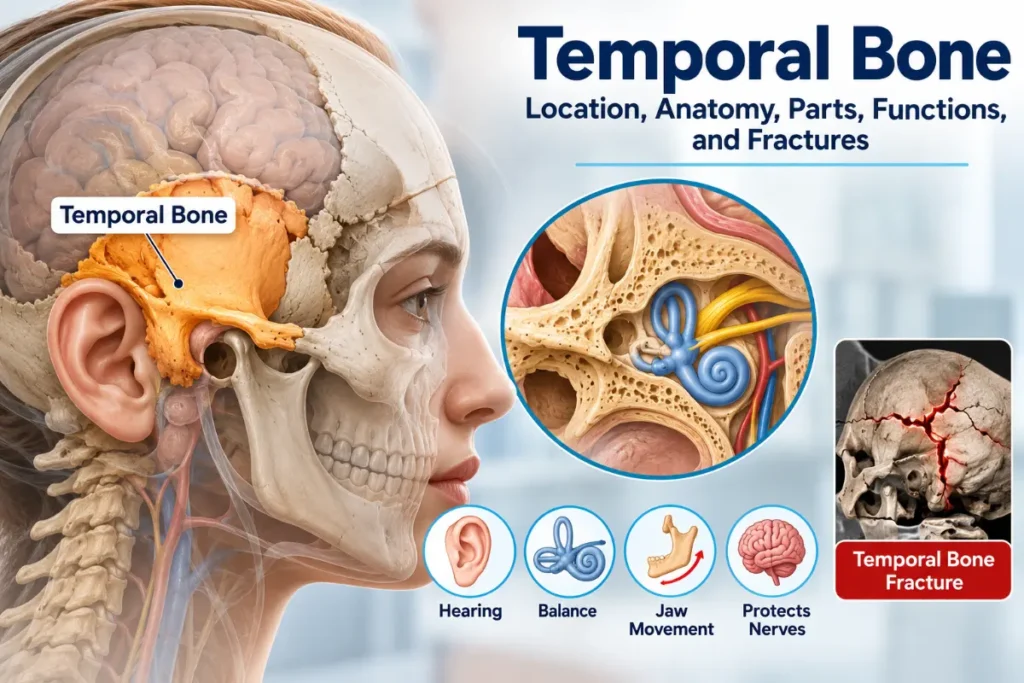
The temporal bone is one of two paired bones located on the sides and base of the human skull, near the ears. Each temporal bone forms part of the cranial base, protects structures involved in hearing and balance, supports jaw movement, and provides pathways for important nerves and blood vessels. Its four major anatomical portions are the squamous, petrous, mastoid, and tympanic parts.
Understanding temporal bone anatomy helps explain how the skull protects the delicate structures of the ear and supports the temporomandibular joint (TMJ). Because several important nerves and sensory structures pass through or lie within this bone, injuries can affect hearing, balance, and facial movement.
Where Is the Temporal Bone Located?
The temporal bones lie on the lower sides of the skull, roughly at ear level. Each extends inward to form part of the skull base and contributes to the middle and posterior cranial fossae, which accommodate portions of the brain.
Each temporal bone connects with five surrounding bones:
- Parietal bone: Forms much of the upper side of the skull.
- Occipital bone: Forms the back and part of the base of the skull.
- Sphenoid bone: Contributes to the central skull base.
- Zygomatic bone: Joins the temporal bone to form the cheek arch.
- Mandible: Articulates with the temporal bone to form the jaw joint.
One of the easiest landmarks to identify is the mastoid process, a rounded bony projection that can be felt just behind the ear.
These relationships help explain the temporal bone’s position within the anatomy of the temporal region and its contribution to the skull’s overall structure.
What Are the Four Parts of the Temporal Bone?
The temporal bone is commonly divided into four major anatomical portions. Although they form a continuous bone, each has distinct features and functions. The anatomical classification of the temporal bone identifies the squamous, petrous, mastoid, and tympanic portions.
1. Squamous Part
The squamous part is the thin, flattened upper region of the temporal bone. It forms part of the lateral skull wall, helping enclose and protect the brain.
Two important structures are associated with this region:
- Zygomatic process: Extends forward to join the zygomatic bone, forming the zygomatic arch.
- Mandibular fossa: A depression that receives the mandibular condyle and contributes to the TMJ.
Together, the mandibular fossa and condyle form part of the joint that allows the jaw to open, close, and move during chewing and speaking.
2. Petrous Part
The petrous part is a dense, pyramid-shaped structure extending inward at the skull base. It contains the inner ear, including the cochlea, vestibule, and semicircular canals. These structures are responsible for detecting sound and sensing head movement.
The petrous portion also contains important passages:
- Internal acoustic meatus: Carries the facial nerve (cranial nerve VII), vestibulocochlear nerve (cranial nerve VIII), and associated blood vessels, typically including the labyrinthine artery.
- Carotid canal: Provides a passage for the internal carotid artery, which supplies blood to the brain.
The petrous portion’s dense structure helps protect the delicate inner ear. Its anatomical arrangement is particularly important when examining hearing loss, balance disorders, and injuries affecting nearby nerves.
3. Mastoid Part
The mastoid part lies behind the ear and contains the mastoid process, a prominent projection that provides attachment points for several muscles.
One of these is the sternocleidomastoid muscle, which helps turn and tilt the head.
Inside the mastoid region are interconnected air-filled spaces called mastoid air cells. These communicate with the middle ear and form part of its connected air-space system.
4. Tympanic Part
The tympanic part forms much of the bony external acoustic meatus, the passage leading toward the eardrum.
It helps create the bony portion of the external ear canal and supports structures near the tympanic membrane.
The external acoustic meatus should not be confused with the internal acoustic meatus. The external passage conducts sound toward the eardrum, while the internal passage carries nerves and blood vessels.
This distinction is important because the external ear canal contains both cartilaginous and bony portions, whereas the internal acoustic meatus lies within the petrous temporal bone.
Summary of the Four Temporal Bone Parts
| Part | Location and Structure | Main Role |
| Squamous | Thin, flattened lateral region | Skull protection and jaw joint formation |
| Petrous | Dense, pyramid-shaped skull-base region | Protects inner ear structures and contains major passages |
| Mastoid | Region behind the ear containing air cells | Muscle attachment and connection with middle ear air spaces |
| Tympanic | Region surrounding the bony ear canal | Forms and supports part of the external acoustic meatus |
Important Landmarks of the Temporal Bone
The styloid process is a thin, pointed projection extending downward from the temporal bone. It provides attachment points for muscles and ligaments associated with the tongue, throat, and hyoid region.
Other important landmarks include the mastoid process behind the ear, the zygomatic process forming part of the cheek arch, and the mandibular fossa involved in jaw movement.
The external and internal acoustic meatuses are also significant anatomical landmarks. Although their names are similar, they have different locations and functions.
Recognizing these structures is useful when studying skull anatomy or interpreting medical imaging of the temporal bone, particularly because some landmarks are not visible from the outside of the skull.
What Are the Functions of the Temporal Bone?
The temporal bone has several important functions, particularly in protecting sensory structures, supporting movement, and providing passageways for nerves and blood vessels.
It surrounds the middle and inner ear, protecting structures that transmit and detect sound. The cochlea converts sound vibrations into nerve signals, while the vestibular apparatus senses head movement and helps the body maintain balance.
The bone also contributes to the TMJ, allowing the lower jaw to move during activities such as chewing and speaking. Its mastoid and styloid processes provide attachment points for muscles and ligaments involved in movements of the head, neck, tongue, and throat.
Several important structures travel through openings and canals within the temporal bone, including the facial and vestibulocochlear nerves and the internal carotid artery.
Although the temporal bone is closely associated with hearing and balance, it does not perform these sensory functions itself. Instead, it houses and protects the organs responsible for them.
Temporal Bone Fractures: Causes, Symptoms, and Warning Signs
A temporal bone fracture is a break in the bone, usually caused by significant head trauma. Motor vehicle collisions, serious falls, and direct blows to the head are common causes.
Because the bone surrounds the ear and several important nerves, a fracture may cause:
- Hearing loss in one or both ears
- Dizziness, vertigo, or problems maintaining balance
- Facial weakness or paralysis
- Bleeding from the ear
- Clear fluid draining from the ear, potentially indicating a cerebrospinal fluid (CSF) leak
- Bruising behind the ear, known as Battle sign, which can develop after certain skull fractures
The symptoms depend on where the fracture occurs and which structures are damaged. Some complications improve with appropriate medical care, while others may cause lasting hearing, balance, or facial nerve problems.
Battle sign and other individual symptoms do not confirm a temporal bone fracture on their own. A medical examination and appropriate imaging are needed to evaluate a suspected injury.
When Is Emergency Medical Care Necessary?
A suspected temporal bone fracture requires emergency medical evaluation, particularly after a serious head injury.
Warning signs include new hearing loss, severe dizziness, facial weakness, blood or clear fluid draining from the ear, confusion, loss of consciousness, or bruising behind the ear after trauma.
Doctors generally use CT imaging to assess a suspected fracture and look for associated injuries. Hearing tests and facial nerve examinations may also be needed, depending on the symptoms.
The clinical evaluation of temporal bone fractures focuses on identifying damage to hearing structures, the vestibular system, the facial nerve, and surrounding tissues.
Prompt medical assessment matters because some complications, including CSF leakage and nerve injury, require specialized care.
Read more: Heating Pad: Benefits, Types, Safe Use, and Limitations
Frequently Asked Questions
1. Is the temporal bone a paired bone?
Yes. The human skull contains two temporal bones, one on each side near the ears. Together, they form parts of the lateral skull walls and cranial base. Each bone has corresponding anatomical regions and helps protect the structures of its respective ear.
2. What organs does the temporal bone protect?
The temporal bone protects structures of the middle and inner ear, including the cochlea, vestibule, and semicircular canals. These structures are essential for hearing and balance. The bone also surrounds important nerves and blood vessels that travel through the skull.
3. What happens if the temporal bone is fractured?
A fracture can injure structures involved in hearing, balance, or facial movement. Symptoms may include hearing loss, dizziness, facial weakness, bleeding, or clear fluid drainage from the ear. The severity depends on the location and extent of the injury. Anyone with a suspected temporal bone fracture after head trauma needs emergency medical evaluation.
4. Can the temporal bone cause jaw pain?
Yes. The temporal bone forms part of the temporomandibular joint (TMJ), which connects the lower jaw to the skull. Problems affecting this joint can cause pain near the ear, jaw stiffness, clicking, or difficulty chewing. However, jaw pain is often related to the joint or surrounding muscles rather than damage to the temporal bone itself.

0 Comments