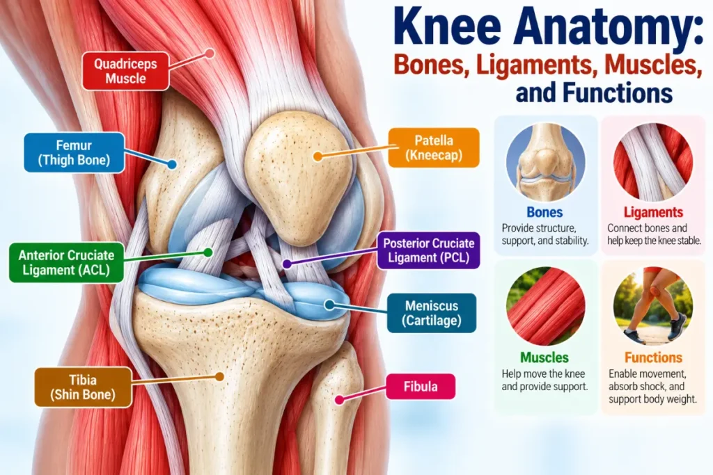
The knee is a modified hinge-type synovial joint that connects the thigh to the lower leg. It consists of three principal bones: the femur (thighbone), tibia (shinbone), and patella (kneecap). The nearby fibula provides attachment points for supporting tissues but is not part of the knee’s main articulations. These are the tibiofemoral joint, where the femur meets the tibia, and the patellofemoral joint, where the patella meets the femur. This arrangement allows the knee to bend, straighten, rotate slightly, and support body weight.
Bones alone cannot keep the knee stable. Ligaments prevent excessive movement, muscles and tendons control motion, and cartilage helps protect the joint surfaces. The menisci also distribute pressure between the femur and tibia. These structures work together whenever you walk, run, climb stairs, or squat.
What Is the Knee Joint?
The knee is the body’s largest joint and one of its most complex. Unlike a simple door hinge, it permits a small amount of rotation in addition to bending and straightening. This flexibility helps the leg adapt to different movements while maintaining stability.
To understand knee anatomy, it helps to know four anatomical directions:
- Anterior: The front of the knee, where the patella sits.
- Posterior: The back of the knee.
- Medial: The inner side, toward the body’s midline.
- Lateral: The outer side, away from the body’s midline.
These terms describe the positions of bones, ligaments, and other tissues within the joint.
Bones of the Knee and Their Functions
Three bones form the knee’s main articulations, while a fourth, the fibula, supports nearby structures. Each has a distinct anatomical role.
Femur (Thighbone)
The femur forms the upper part of the knee. Its lower end has two rounded projections called femoral condyles, which articulate with the tibia. A groove on the front of the femur guides the patella as the knee bends and straightens.
Tibia (Shinbone)
The tibia is the larger, weight-bearing bone of the lower leg. Its broad upper surface, known as the tibial plateau, supports the femoral condyles. The menisci lie between these surfaces, helping distribute the forces passing through the joint.
Patella (Kneecap)
The patella is a small, triangular bone embedded in the quadriceps tendon at the front of the knee. It provides some protection for the joint and improves the quadriceps’ mechanical advantage, making knee extension more efficient.
Fibula
The fibula is the slender bone running along the outer side of the lower leg. Its upper end, called the fibular head, provides attachment points for the lateral collateral ligament and several muscles. Although important for supporting the knee, the fibula does not form part of its principal weight-bearing articulation.
Knee Ligaments: ACL, PCL, MCL, and LCL
Ligaments are strong bands of connective tissue that connect bones and help prevent excessive joint movement. The knee has four principal stabilizing ligaments, each controlling movement in particular directions.
| Ligament | Location | Primary function |
| Anterior cruciate ligament (ACL) | Inside the knee, crossing the PCL | Limits forward movement of the tibia and helps control rotation |
| Posterior cruciate ligament (PCL) | Inside the knee, behind the ACL | Limits backward movement of the tibia |
| Medial collateral ligament (MCL) | Inner side of the knee | Resists excessive inward angulation (valgus stress) |
| Lateral collateral ligament (LCL) | Outer side of the knee | Resists excessive outward angulation (varus stress) |
Cruciate Ligaments (ACL and PCL)
The ACL and PCL cross in an X-shaped arrangement near the center of the knee. Both lie within the fibrous joint capsule but outside the synovial cavity.
Their positions help control forward and backward movement between the femur and tibia. The ACL is particularly important for rotational control, while the PCL provides substantial resistance to backward displacement of the tibia.
Collateral Ligaments (MCL and LCL)
The MCL runs along the inner knee, connecting the femur to the tibia. The LCL extends along the outer knee, connecting the femur to the fibular head.
These ligaments resist excessive side-to-side angulation and help maintain alignment, especially when the knee is straight.
Knee Cartilage and Menisci
The knee contains two types of cartilage with different structures and purposes. Both are essential for managing the forces generated during movement.
Articular Cartilage
Articular cartilage is a smooth layer of hyaline cartilage covering the joint surfaces of the femur, tibia, and patella. It helps the bones glide past one another with very little friction while distributing pressure across the joint.
Unlike many other tissues, articular cartilage has a limited ability to repair itself after significant damage. Its smooth surface is therefore important for normal knee function.
Medial and Lateral Menisci
The menisci are two crescent-shaped pads of fibrocartilage between the femur and tibia.
- Medial meniscus: Located on the inner side of the knee. Its attachments make it less mobile than the lateral meniscus.
- Lateral meniscus: Located on the outer side. It generally moves more freely as the knee bends and rotates.
Their wedge-shaped structure improves contact between the rounded femoral condyles and the tibial plateau. By spreading forces over a larger area, the menisci help absorb shock, distribute weight, and improve stability.
The distinction is important: articular cartilage covers bone surfaces, while the menisci sit between those surfaces to help manage pressure. Both are described in more detail in this overview of knee cartilage and joint structures.
Knee Tendons and Supporting Muscles
Tendons attach muscles to bones and transmit the force needed to move the joint. Ligaments, by comparison, connect bones to bones and primarily help maintain stability.
Quadriceps Tendon and Patellar Tendon
The quadriceps tendon connects the quadriceps muscle group to the patella. Below the kneecap, the patellar tendon, also known as the patellar ligament, attaches the patella to the tibial tuberosity.
Together, these structures form part of the extensor mechanism. When the quadriceps contract, force passes through this system to straighten the knee.
Muscles That Move the Knee
Several muscles cross or act on the knee, but four groups have particularly important roles:
- Quadriceps: Located at the front of the thigh, these muscles are responsible for most knee extension.
- Hamstrings: Found at the back of the thigh, they bend the knee and help control rotation.
- Popliteus: A small muscle behind the knee that helps initiate flexion from the fully extended, locked position.
- Gastrocnemius: A major calf muscle that crosses the back of the knee and assists with flexion.
These muscles do more than produce movement. By controlling joint position and responding to changing loads, they provide dynamic stability during everyday activities.
Joint Capsule, Synovial Membrane, and Bursae
Several softer tissues surround the knee and allow its structures to move without excessive friction.
Joint capsule: A tough fibrous enclosure surrounding the joint. It provides support and encloses the synovial lining.
Synovial membrane: This tissue lines the inner surface of the fibrous capsule and forms folds and reflections inside the joint. It does not cover articular cartilage or the free surfaces of the menisci. The membrane produces synovial fluid, which lubricates the joint and helps nourish cartilage.
Bursae: Small, fluid-filled sacs positioned where tissues might otherwise rub against one another. Important examples include the suprapatellar, prepatellar, and infrapatellar bursae.
The placement and functions of these structures are described in this anatomical explanation of the knee joint.
How Does the Knee Move and Stay Stable?
The knee’s movements depend on coordinated activity between muscles, bones, ligaments, and cartilage.
Flexion is the bending movement that brings the lower leg toward the thigh. The hamstrings are among the main muscles responsible.
Extension straightens the knee, mainly through quadriceps contraction and the extensor mechanism.
Rotation involves limited inward and outward turning of the tibia relative to the femur. It occurs primarily when the knee is flexed, rather than fully straightened.
Walking requires the knee to alternate smoothly between flexion and extension while carrying body weight. Running and squatting place different demands on the joint, requiring greater muscular control and coordinated force distribution.
The ligaments and joint capsule provide passive stability by restricting excessive motion. Muscles supply active control, while the menisci increase contact between the femur and tibia. Articular cartilage allows the joint surfaces to glide smoothly under load.
Consider standing up from a chair. The quadriceps straighten the knees, surrounding muscles help maintain alignment, and the ligaments prevent excessive joint movement. Meanwhile, the cartilage and menisci distribute pressure as body weight shifts onto the legs.
This coordination explains how the knee combines mobility with the stability needed for weight-bearing. A broader knee joint anatomy overview also explains how its components contribute to movement and balance.
Common Knee Injuries and the Structures They Affect
Knowing which structures support the knee helps explain why different injuries can affect its movement and stability.
ACL tears damage a major stabilizing ligament that limits forward tibial movement and helps control rotation. These injuries may occur during sudden changes in direction, pivoting, or awkward landings.
Meniscus tears affect the fibrocartilage pads responsible for distributing weight between the femur and tibia. Twisting injuries and age-related tissue changes are common causes. The medical guidance on meniscus tears explains the structures involved and how these injuries develop.
Collateral ligament injuries damage the MCL or LCL. Because these ligaments resist excessive side-to-side angulation, an injury can reduce stability in the affected direction.
Knee osteoarthritis is a joint disease involving deterioration of articular cartilage along with changes in underlying bone and other joint tissues. It affects more than cartilage alone, as explained in this knee osteoarthritis overview.
These conditions demonstrate how damage to a particular structure can interfere with the knee’s normal mechanics.
Frequently Asked Questions
1. What are the main parts of the knee?
The knee’s main structures include the femur, tibia, patella, ligaments, tendons, menisci, articular cartilage, muscles, and joint capsule. The nearby fibula also provides important attachment points. Each contributes to movement, weight-bearing, or stability.
2. What are the four main ligaments of the knee?
The knee’s main structures include the femur, tibia, patella, ligaments, tendons, menisci, articular cartilage, muscles, and joint capsule. The nearby fibula also provides important attachment points. Each contributes to movement, weight-bearing, or stability.
3. What is the difference between knee cartilage and meniscus?
Articular cartilage is smooth hyaline cartilage covering the bone surfaces within the knee. It reduces friction and helps distribute pressure. The menisci are two crescent-shaped fibrocartilage pads between the femur and tibia that absorb shock, spread weight, and improve joint stability.
4. Which muscles control knee movement?
The quadriceps primarily straighten the knee, while the hamstrings bend it and help control rotation. The popliteus assists in initiating flexion from full extension, and the gastrocnemius contributes to bending. These muscles also help keep the joint stable during movement.
5. Why is the knee considered a modified hinge joint?
The knee is classified as a modified hinge joint because it mainly bends and straightens but also allows a limited amount of rotation, especially when flexed. This combination makes movements such as walking, running, turning, and squatting possible.

0 Comments