Transitional epithelium, also called urothelium, is a specialized type of stratified epithelial tissue that lines parts of the urinary tract, including the renal calyces, renal pelvis, ureters, and urinary bladder. Its two main functions are allowing urinary organs to stretch as they fill with urine and protecting the underlying tissues from substances in urine. Unlike most epithelial tissues, its superficial cells can change shape as these organs expand and contract.
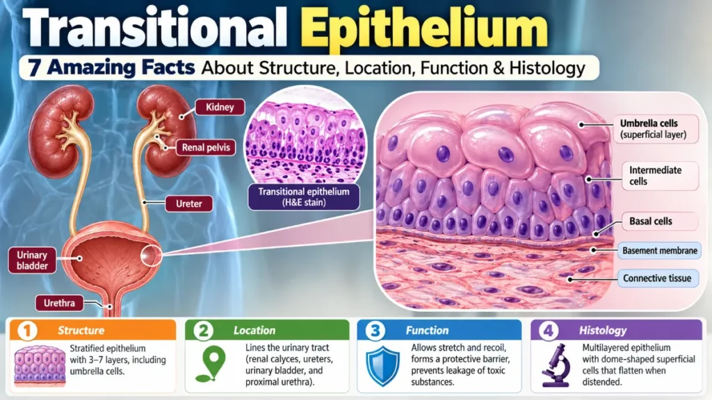
Large, rounded cells called umbrella cells cover the tissue’s inner surface. When the bladder is empty, these cells usually appear dome-shaped. As the bladder fills, they flatten and spread out, helping the lining expand while preserving its protective barrier. This ability to change appearance is also one of the features that makes transitional epithelium recognizable under a microscope.
Where Is Transitional Epithelium Found?
Transitional epithelium is found primarily in parts of the urinary system responsible for collecting, transporting, and storing urine. Because these structures experience changes in volume, their lining needs to remain flexible without allowing urine to pass freely into surrounding tissues.
The main transitional epithelium locations include:
- Renal calyces: Cup-shaped structures that collect urine from the renal papillae.
- Renal pelvis: A funnel-shaped region that collects urine and directs it into the ureter.
- Ureters: Muscular tubes that carry urine from the kidneys to the bladder.
- Urinary bladder: A hollow muscular organ that stores urine until urination.
- Parts of the urethra: The proximal urethra contains urothelium, while other portions have different epithelial linings.
The urinary bladder provides a particularly clear example of this tissue’s flexibility. As the bladder fills and empties, its epithelial lining changes appearance to accommodate the changing volume.
Structure of Transitional Epithelium
The transitional epithelium structure consists of several layers of cells resting on a basement membrane. Its thickness and appearance vary depending on the location and how much the tissue is stretched.
According to the histological description of the urinary bladder, the urothelium has three main cellular zones: basal, intermediate, and superficial.
Basal, Intermediate, and Umbrella Cells
1. Basal cells
Basal cells form the deepest epithelial layer and rest directly on the basement membrane. They are generally small and cuboidal, although their shapes can vary.
These cells help maintain and renew the epithelial lining, replacing cells as part of the tissue’s normal repair and regeneration processes.
2. Intermediate cells
Intermediate cells occupy the region between the basal and superficial layers. They are usually polygonal or pear-shaped and make up much of the tissue’s thickness.
Their arrangement allows the lining to adjust as the urinary organ expands or returns to its relaxed state.
3. Superficial umbrella cells
Umbrella cells are large, specialized cells that cover the surface exposed to urine. In relaxed tissue, they usually have a rounded or dome-shaped appearance.
Some umbrella cells contain two nuclei, a feature called binucleation. This can develop during cellular differentiation, although the underlying processes vary. Not every umbrella cell has two nuclei.
Their specialized membranes and tight connections with neighboring cells are especially important for preventing urine constituents from passing into deeper tissues.
What Is the Function of Transitional Epithelium?
Transitional epithelium has two essential functions: allowing urinary organs to expand and forming a protective barrier against urine.
Stretching and expansion
The urinary bladder must accommodate increasing amounts of urine without damaging its lining.
As the bladder fills, the urothelial cells change shape, and the epithelial surface expands. Changes in the membrane organization of umbrella cells also help the lining adapt to stretching.
This flexibility allows the bladder to store urine while preserving the integrity of its epithelial barrier.
Protection against urine
Urine contains dissolved salts, metabolic waste products, and other substances that could irritate or damage tissues if they passed through the lining.
The urothelium limits this movement through several specialized features:
- Tight junctions: Restrict the passage of substances between neighboring cells.
- Uroplakin proteins: Form protective plaques on the surface membranes of umbrella cells.
- Specialized apical membranes: Help prevent urine constituents from penetrating the epithelial barrier.
These features work together to create a highly resistant lining. Research on the urothelial barrier and its protective mechanisms explains how surface proteins and tight junctions contribute to this function.
How Transitional Epithelium Changes When Stretched
One of the most distinctive features of this tissue is how its microscopic appearance changes when the urinary bladder fills.
When the bladder is empty:
The epithelial lining looks relatively thick, and the superficial umbrella cells appear rounded or dome-shaped. The bladder’s mucosal surface also commonly forms folds.
When the bladder is full:
The umbrella cells flatten and spread across a larger surface area. The lining becomes thinner in appearance, and fewer cellular layers may be distinguishable in a tissue section.
These changes are normally reversible. As the bladder empties, the lining can return toward its relaxed appearance.
The bladder histology explanation from the University of Leeds also describes how the folded lining accommodates considerable changes in bladder volume.
Although stretched transitional epithelium may resemble stratified squamous epithelium, it does not actually transform into a different tissue type.
How to Identify Transitional Epithelium Under a Microscope
Transitional epithelium histology is commonly studied using stained tissue sections from the urinary bladder or ureter. Its multiple cellular layers and distinctive umbrella cells make it relatively easy to recognize once you know what to look for.
Start by identifying the lumen, the open space through which urine passes or collects. Next, examine the cells lining that space and the smaller cells beneath them.
Look for these five features:
- Multiple cell layers: Several layers of epithelial cells are present rather than a single row.
- Large surface cells: Umbrella cells are generally larger than the cells beneath them.
- Rounded surface contours: In relaxed tissue, umbrella cells often project toward the lumen with dome-shaped surfaces.
- Occasional binucleated cells: Some superficial cells contain two visible nuclei.
- Underlying connective tissue: A basement membrane separates the epithelium from the connective tissue beneath it.
A relaxed bladder specimen may show approximately five to seven apparent cellular layers, while stretched tissue may appear to contain only two or three. These counts are approximate because the appearance depends on tissue location, distension, and how the specimen was sectioned.
The human bladder histology material describes the dome-shaped superficial cells seen in contracted bladder tissue.
For reliable identification, consider the overall arrangement rather than relying on a single feature. The combination of multiple layers, large superficial cells, and changes in surface shape is especially characteristic of urothelium.
Transitional Epithelium vs. Other Epithelial Tissues
Transitional epithelium can look similar to other epithelial tissues, particularly when stretched. However, differences in cell organization, surface appearance, and function make identification easier.
| Feature | Transitional Epithelium | Stratified Squamous Epithelium | Simple Cuboidal Epithelium |
| Cell layers | Multiple layers | Multiple layers | Single layer |
| Surface cell shape | Dome-shaped when relaxed; flatter when stretched | Flattened | Cuboidal |
| Main function | Stretching and urinary protection | Protection against abrasion, as in the esophagus | Absorption and secretion |
| Typical location | Urinary bladder | Esophagus | Kidney tubules |
| Distinctive feature | Large umbrella cells | Flattened superficial cells | One layer of cube-shaped cells |
The main difference between transitional and stratified squamous epithelium lies in their superficial cells and specialized functions.
Stratified squamous epithelium has flattened surface cells that help protect against abrasion. Transitional epithelium, by contrast, has umbrella cells that change shape during stretching while maintaining a barrier against urine.
Simple cuboidal epithelium is easier to distinguish because it contains only one layer of cube-shaped cells.
When studying histology slides, remember that large superficial umbrella cells and a multilayered arrangement are among the clearest clues that you are looking at urothelium.
Read also: Ronnie Coleman Now: 2026 Health Update, Mobility, and Life Today
Frequently Asked Questions
1. Why is transitional epithelium called transitional?
It is called transitional because the shape and arrangement of its cells appear to change as urinary organs stretch and relax. Its surface cells look rounded when relaxed and flatter when stretched. Despite these changes, the tissue remains the same specialized type of epithelium.
2. Why do some umbrella cells have two nuclei?
Some umbrella cells develop two nuclei during cellular differentiation. One process that can contribute is nuclear division without complete separation of the cell. However, umbrella-cell development involves several cellular mechanisms, and not all umbrella cells are binucleated.
3. Is transitional epithelium present in the kidneys?
Yes. Transitional epithelium lines the renal calyces and renal pelvis, where urine collects before moving into the ureters. However, most kidney tubules have different epithelial linings, including simple cuboidal epithelium.
4. How many layers does transitional epithelium have?
The apparent number of layers depends on how much the tissue is stretched. A relaxed bladder lining may show approximately five to seven layers, while a stretched lining may appear to have only two or three. These numbers can vary with the specimen and anatomical location.
5. What happens if the urothelial barrier is damaged?
A damaged urothelial barrier may allow more urine constituents to reach underlying tissues, potentially causing irritation or inflammation. The effects depend on the location, extent, and cause of the damage. Maintaining an effective barrier is therefore essential for normal urinary tract function.


0 Comments