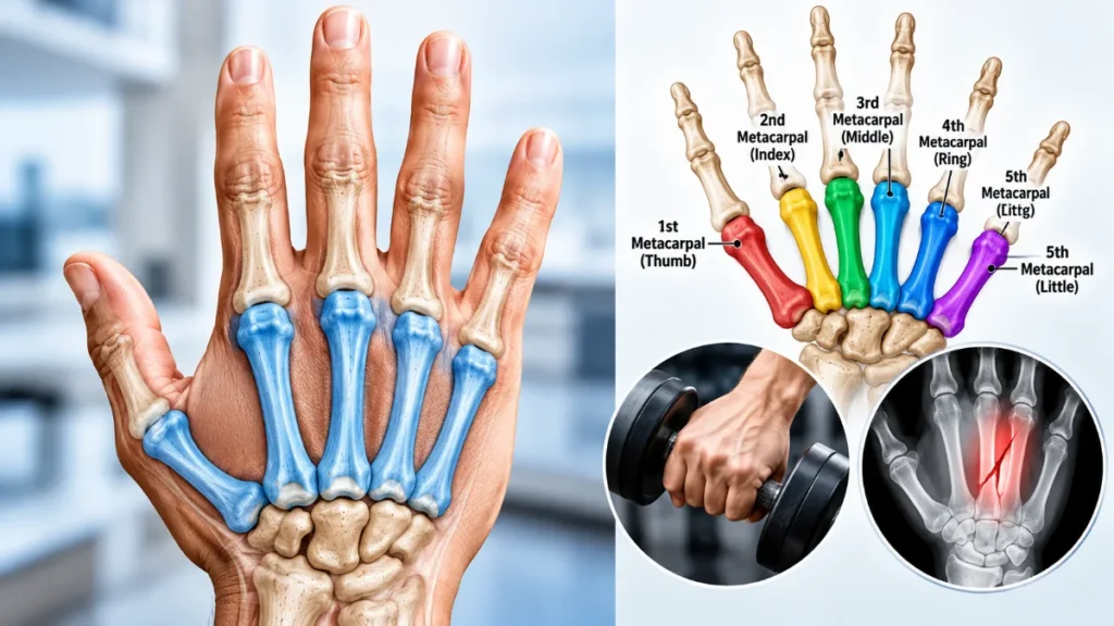
The metacarpals are five long bones that form the framework of the palm of each hand. Located between the wrist and fingers, these bones are numbered I through V, starting at the thumb and ending at the little finger. They support the palm, connect the wrist to the fingers, and help with everyday movements such as gripping, grasping, and holding objects.
Each hand contains eight carpal bones in the wrist, five metacarpal bones in the palm, and 14 phalanges in the fingers and thumb. These bones work together, but each group has a distinct role. Understanding the metacarpal bones and their anatomy helps explain how the hand moves and why injuries to the palm can affect grip strength and finger movement.
Where Are the Metacarpals Located?
The metacarpal bones lie in the palm, extending from the carpal bones of the wrist toward the proximal phalanges, the first bones of the fingers and thumb.
Each metacarpal corresponds to a particular digit:
- First metacarpal (I): Located at the base of the thumb.
- Second metacarpal (II): Extends toward the index finger.
- Third metacarpal (III): Aligns with the middle finger.
- Fourth metacarpal (IV): Aligns with the ring finger.
- Fifth metacarpal (V): Aligns with the little finger.
The rounded ends of these bones form part of the knuckles you see when making a fist. Together, the metacarpals give the palm its shape and provide a stable foundation for finger movement.
Anatomy of the Five Metacarpal Bones
Metacarpals are classified as long bones because they have an elongated central shaft and enlarged ends. Although all five share the same basic structure, their shapes and mobility vary depending on their positions in the hand.
Parts of a Metacarpal Bone
A typical metacarpal has four identifiable regions:
- Base: The proximal end, nearest the wrist, which connects with the carpal bones. The bases of metacarpals II–V also form joints with their adjacent metacarpals.
- Shaft (body): The long central portion that provides structural support and attachment sites for muscles.
- Neck: The narrower area between the shaft and head, where certain fractures commonly occur.
- Head: The rounded distal end that meets the proximal phalanx to form a knuckle joint.
The first metacarpal is shorter and more mobile than the others, giving the thumb greater freedom of movement.
By comparison, the second and third metacarpals are relatively stable, providing a firm foundation for gripping. The fourth and fifth can move more freely at their bases, helping the palm curve around objects.
What Bones and Joints Connect to the Metacarpals?
The metacarpals connect to the wrist bones, finger bones, and neighboring metacarpals through several joints. These connections provide a balance of stability and flexibility, allowing the hand to perform both powerful grips and precise movements.
Carpometacarpal (CMC) Joints
The carpometacarpal joints connect the bases of the metacarpals to specific carpal bones.
| Metacarpal | Corresponding Digit | Carpal Bones It Articulates With |
| First (I) | Thumb | Trapezium |
| Second (II) | Index finger | Trapezium, trapezoid, capitate |
| Third (III) | Middle finger | Capitate |
| Fourth (IV) | Ring finger | Capitate, hamate |
| Fifth (V) | Little finger | Hamate |
The second and third CMC joints allow relatively little movement, making the central part of the hand more stable. The fourth and fifth joints are more flexible, helping the palm adjust when grasping objects.
Metacarpophalangeal (MCP) Joints
The MCP joints connect the rounded heads of the metacarpals to the bases of the proximal phalanges.
Commonly known as the knuckle joints, they allow the fingers to bend, straighten, spread apart, and move together. The thumb’s MCP joint mainly permits bending and straightening, with more limited side-to-side movement.
Intermetacarpal Joints
The bases of the second through fifth metacarpals form joints with their respective neighboring metacarpals.
These small joints help keep the palm stable while permitting limited movement. Together with the CMC joints, they allow the hand to adjust its shape during gripping and other activities.
Why Is the First Metacarpal Different?
The first metacarpal forms a saddle-shaped CMC joint with the trapezium, one of the wrist bones. Unlike the relatively rigid joints near the index and middle fingers, this joint permits movement in multiple directions.
This mobility makes thumb opposition possible. During opposition, the thumb moves across the palm toward the other fingertips, allowing you to pinch, pick up small objects, and handle items precisely.
These movements rely on coordinated action between the joint, surrounding muscles, ligaments, and other soft tissues.
What Are the Functions of the Metacarpals?
The metacarpal bones provide the framework that supports many everyday hand movements. Their arrangement helps the hand remain stable while adjusting to objects of different shapes and sizes.
Their main functions include:
- Supporting the palm: They maintain the palm’s shape and provide attachment points for muscles and connective tissues.
- Supporting gripping and grasping: They help position the fingers and transfer forces when holding, squeezing, or wrapping the hand around objects.
- Facilitating finger movement: Through the MCP joints, they support bending, straightening, and side-to-side finger movements.
- Supporting thumb mobility: The first metacarpal helps make pinching, opposition, and precise object handling possible.
- Distributing forces: They help transfer mechanical loads between the fingers and wrist during gripping and weight-bearing activities.
Although these bones provide essential structural support, they do not move the hand independently. Muscles, tendons, ligaments, and nerves work together with the metacarpals to produce controlled movement.
Common Metacarpal Injuries and Fractures
Metacarpal fractures are common hand injuries that can result from falls, direct blows, sports injuries, or other trauma. A fracture may occur at the base, shaft, neck, or head of the bone.
The location and severity of the break can affect finger alignment, joint movement, and grip strength.
Types and Symptoms of Metacarpal Fractures
Two important examples are:
- Boxer’s fracture: Usually involves the neck of the fifth metacarpal, often after someone strikes a hard object with a clenched fist.
- Thumb-base fracture: Affects the base of the first metacarpal. Certain injuries, including Bennett and Rolando fractures, extend into the thumb’s CMC joint and may affect its stability.
According to clinical guidance on hand fractures, common symptoms include pain, swelling, bruising, tenderness, difficulty moving the affected finger, and reduced hand function.
More severe fractures can cause visible deformity, a depressed knuckle, or abnormal finger alignment. Sometimes, the injured finger crosses over a neighboring finger when the person attempts to make a fist.
Fractures involving the base of the thumb deserve particular attention because damage to the joint surface can interfere with thumb movement and stability.
How Are Metacarpal Fractures Diagnosed and Treated?
A healthcare professional typically examines the injured hand for swelling, tenderness, deformity, finger alignment, movement, and circulation. X-rays are usually used to locate the fracture and determine whether the bone fragments have shifted.
Treatment depends on the fracture’s location, alignment, stability, and severity.
Common approaches include:
- Immobilization: A splint or cast holds a stable, acceptably aligned fracture in position while it heals.
- Closed reduction: A healthcare professional repositions displaced bone fragments without making a surgical incision.
- Surgery: Some unstable, significantly displaced, rotated, or joint-involving fractures require surgical fixation to restore proper alignment.
Once healing has progressed sufficiently, a healthcare professional may recommend rehabilitation exercises to help restore flexibility, movement, and hand function.
Anyone with a suspected metacarpal fracture should seek medical assessment. Visible deformity, numbness, an open wound, or signs of poor circulation require urgent evaluation.
Read also: Arm Bones: Anatomy, Names, Locations, and Functions
Frequently Asked Questions
What Is the Difference Between Metacarpals and Metatarsals?
Metacarpals are the five long bones that form the framework of each palm, while metatarsals are the five long bones in each foot, located between the tarsal bones and toe phalanges. Both groups provide skeletal support, but their functions differ. Metacarpals help with gripping, finger positioning, and hand movement, whereas metatarsals support standing, balance, and walking.

0 Comments